A white blood cell (WBC) count is a test that measures the number of white blood cells in your body. This test is often included with a complete blood count (CBC). The term “white blood cell count” is also used more generally to refer to the number of white blood cells in your body.
There are several types of white blood cells, and your blood usually contains a percentage of each type. Sometimes, however, your white blood cell count can fall or rise out of the healthy range.

Also Known As: WBC Count, Leukocyte Count, White Count, TLC, Leukocyte
Test Panel: Hemoglobin, Red Blood Cells (RBC), HCT, MCV, MCH, MCHC, Platelets Count, White Blood Cells (WBC), DLC, ESR
Test Requirements
- Patient Sample (EDTA)
- Test Tubes
- Microscope
- Neubauer Counting Chamber
- Micropipettes
- Cover Glasses (Cover Slips)
- Hand counter
- Turk’s Solution
Procedure:
- Take 0.38 ml of Turk’s Colution and 20ul of Blood in a test tube.
- Mix the blood and Turk’s Solution by shaking with thumb and finger over the end.
- Fill The Neubauer Chamber by Micropipette.
- Place the counting chamber on the microscope stage and allow 2-3 min. for the cells to settle.
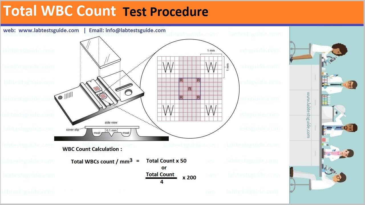
- Count the number of white cells in four large corner squares marked “W”.
- Cells which are touching the left-hand lines or upper lines of the square are included in the count, while cells touching the lower and right margins are excluded.
- After The Countion celles then calculate the total wbc count for this formula
Total WBC Count = Total Cells(4 squares) x 50

Total WBC Count Calculations:
- Count the cells in the Neubauer chamber. These are counted in the four large corner squares labeled as WBC and if the number is Y.
- One large area is 1 x 1 mm, and the depth is 0.1 mm.
- Total area counted in 4 large squares = 4 x 1 x o.1 = 0.4 µL (4/10).
- Y x 10/4 is the total WBC in the cell in 1 µL.
- Now dilution is 1:20.
- Number of WBC in 1µL = Y x 10 x 20/4 = Y x 50 = Total WBC count.
- Total TLC = counted cells (Y) x 50 = TLC/cmm.
- If the count is low <4000/cmm, then use the dilution of 1:10.
- The Source of errors are:
- If there are microclots in the sample.
- If inadequate mixing is done.
- Improper filling of the chamber.
- If the dilutions are improper.
- Mistakes in the calculations.
Referance Ranges:
| Test Name | Male | Female |
| WBC (TLC) | 4.0 – 11.0 x 10⁹ /l | 4.0 – 11.0 x 10⁹ /l |
Types and Identifications of WBC :
The shortest and simplest way to identify white blood cells (WBCs) is by using a microscope to examine a stained blood smear. The most commonly used stain for this purpose is the Wright-Giemsa stain, which differentially stains the various components of blood cells, making WBCs easily distinguishable.
Here are the steps for a rapid identification of WBCs:
- Prepare a Blood Smear: Spread a drop of blood thinly on a glass slide and let it air dry.
- Stain the Smear: Apply Wright-Giemsa stain to the smear. This will typically involve flooding the slide with the stain, followed by a buffer solution, and allowing it to sit for a few minutes.
- Rinse and Dry: Rinse the slide gently with water to remove excess stain and let it air dry.
- Examine Under Microscope: Using a microscope, focus on the stained smear. White blood cells will appear stained and can be distinguished from red blood cells and platelets.
Identification Under Microscope
- Neutrophils: Multi-lobed nucleus with granules in the cytoplasm.
- Lymphocytes: Large, round nucleus with a thin rim of cytoplasm.
- Monocytes: Kidney-shaped or folded nucleus with abundant cytoplasm.
- Eosinophils: Bi-lobed nucleus with large, red-orange granules.
- Basophils: Bi-lobed or S-shaped nucleus with large, dark purple granules.
This method provides a quick and effective way to identify and differentiate between the types of white blood cells.
Possible References Used


