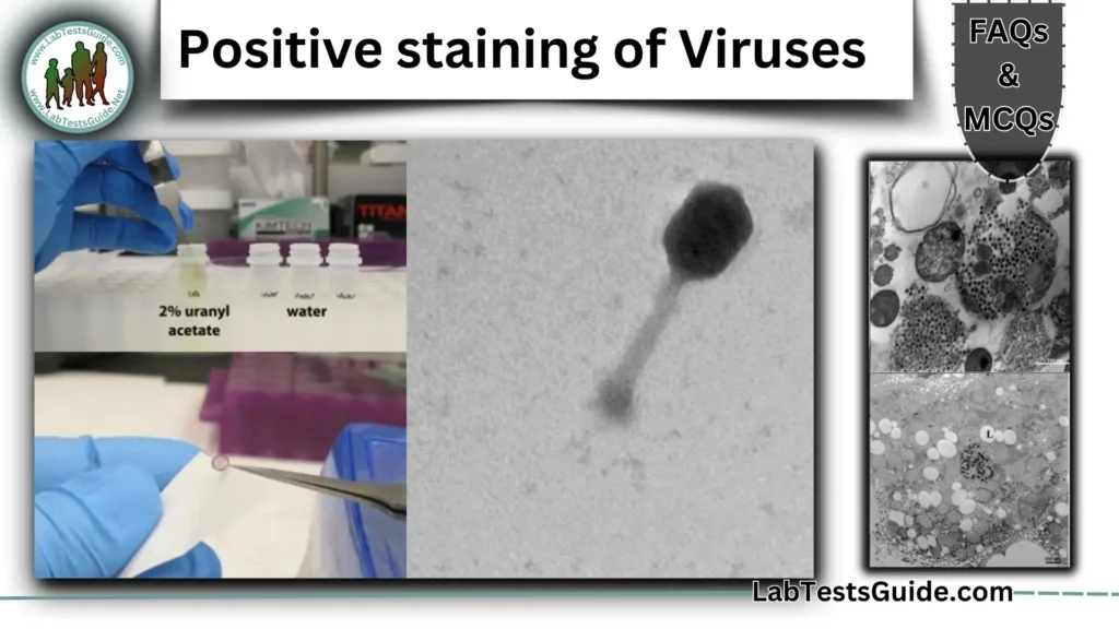
Positive staining of Viruses 50 FAQs and 30 MCQs

Positive staining of Viruses 50 FAQs
What is positive staining of viruses?
Positive staining is an electron microscopy technique where viral particles appear dark against a light background due to heavy metal staining.
How does positive staining differ from negative staining?
In positive staining, viruses appear dark on a light background, whereas in negative staining, they appear light on a dark background.
What is the principle behind positive staining?
Heavy metal salts (e.g., uranyl acetate, lead citrate) bind to viral structures, increasing electron density for better contrast under TEM.
Why is positive staining useful in virology?
It helps visualize viral morphology, including capsids, envelopes, and spikes.
Which viruses can be studied using positive staining?
Enveloped (e.g., HIV, influenza) and non-enveloped (e.g., adenovirus, rotavirus) viruses.
What are the common stains used in positive staining?
Uranyl acetate, lead citrate, and osmium tetroxide.
Why is uranyl acetate used in positive staining?
It binds to proteins and nucleic acids, enhancing contrast.
What is the role of lead citrate in positive staining?
It improves contrast by binding to lipids and proteins.
How is 2% uranyl acetate prepared?
0.04g uranyl acetate dissolved in 2mL ultra-purified water.
Why must uranyl acetate be filtered before use?
To remove undissolved particles that may cause artifacts.
Why is osmium tetroxide used for staining virus envelopes?
It reacts strongly with lipids, highlighting envelope structures.
Is positive staining light-sensitive?
Yes, uranyl acetate degrades under light, so staining should be done in a dark environment.
What are the key steps in positive staining?
Sample preparation, fixation, staining, washing, drying, and TEM observation.
Why is fixation important in positive staining?
It preserves viral structure (e.g., using glutaraldehyde or paraformaldehyde).
How long should a grid be incubated in uranyl acetate?
Typically 15 minutes for uranyl acetate alone, or 30 seconds for quick staining.
Why is washing critical after staining?
Removes excess salts that can distort the image.
How are grids dried after staining?
Air-dried or blotted with filter paper before TEM observation.
Why is CO₂ harmful during lead citrate staining?
CO₂ reacts with lead citrate, forming precipitates that obscure the sample.
How can CO₂ be minimized during staining?
By using NaOH pellets to absorb CO₂ in the environment.
What is the Tokuyasu staining procedure (TSP)?
A positive staining method using uranyl acetate, glutaraldehyde, and polyvinyl alcohol for enhanced virus visualization.
What microscope is used for positive staining?
Transmission Electron Microscope (TEM).
What magnification is used to observe stained viruses?
Typically 10,000x–100,000x.
What does a positively stained virus look like under TEM?
Dark viral particles against a light background.
How does positive staining reveal viral spikes?
Heavy metals bind to glycoproteins, making spikes visible.
Can positive staining show viral nucleic acid cores?
Yes, uranyl acetate binds to nucleic acids, highlighting dense core regions.
What are the advantages of positive staining?
Simple, rapid, high contrast, and useful for structural studies.
What are the limitations of positive staining?
Sensitive to CO₂, light exposure, and may introduce artifacts.
How can staining artifacts be minimized?
Proper washing, avoiding contamination, and controlled staining time.
Does positive staining damage viral structures?
Heavy metals and electron beams can sometimes degrade delicate structures.
Is positive staining expensive?
Requires TEM access and trained personnel, making it costly.
Which viruses have been studied using positive staining?
Hepatitis B, HIV-1, influenza, rotavirus, rubella, and adenoviruses.
How does positive staining help in virus identification?
Reveals unique morphological features (e.g., capsid symmetry, envelopes).
Can positive staining differentiate animal and human viruses?
Yes, by comparing structural features like spikes and envelopes.
How has positive staining contributed to vaccine research?
By providing detailed images of viral structures for antigen design.
What novel virus features have been discovered via positive staining?
E.g., unique rotavirus and rubella virus structures.
Why might a stained virus sample appear blurry?
Due to excess stain, improper washing, or CO₂ contamination.
How can uneven staining be avoided?
Ensure uniform staining time and proper blotting.
What causes salt precipitates on grids?
Inadequate washing or CO₂ exposure during lead citrate staining.
How should grids be stored after staining?
In a grid box, dried overnight before TEM observation.
Can positive staining be automated?
Manual staining is common, but automated systems are being developed.
How does Tokuyasu staining differ from standard positive staining?
It combines multiple reagents (PVA, glutaraldehyde) for better preservation.
Is cryo-EM better than positive staining?
Cryo-EM preserves native structure better but is more complex and expensive.
Can positive staining be combined with immunogold labeling?
Yes, for targeted visualization of specific viral proteins.
How does positive staining compare to negative staining in virus studies?
Positive staining shows internal structures, while negative staining highlights outlines.
What future improvements are expected in positive staining?
New stains, reduced artifacts, and faster protocols.
Is uranyl acetate hazardous?
Yes, it is radioactive and toxic; use PPE (gloves, mask, lab coat).
How should uranyl acetate waste be disposed of?
In designated radioactive/chemical waste containers.
Why is respiratory protection needed during staining?
Uranyl acetate particles can be inhaled.
Can lead citrate be substituted with safer alternatives?
Research is ongoing, but currently, it remains a standard stain.
What safety precautions are needed for osmium tetroxide?
Use in a fume hood; it is highly toxic and volatile.
Positive staining of Viruses 30 MCQs:
- What is the primary purpose of positive staining in electron microscopy?
a) To make the background dark
b) To enhance the contrast of virus particles✔
c) To dissolve viral structures
d) To reduce electron density - How does positive staining differ from negative staining?
a) Positive staining makes viruses appear light on a dark background
b) Positive staining makes viruses appear dark on a light background✔
c) Both techniques produce the same results
d) Negative staining is not used in virology - Which of the following is NOT a common stain used in positive staining?
a) Uranyl acetate
b) Lead citrate
c) Methylene blue✔
d) Osmium tetroxide - What is the role of heavy metal salts in positive staining?
a) They dissolve viral proteins
b) They increase electron density for better contrast✔
c) They make viruses invisible under TEM
d) They prevent staining artifacts
- Why is uranyl acetate commonly used in positive staining?
a) It binds to lipids only
b) It binds to proteins and nucleic acids✔
c) It fluoresces under light
d) It dissolves viral envelopes - What is the correct concentration of uranyl acetate typically used?
a) 0.1%
b) 1%
c) 2%✔
d) 10% - Why must lead citrate staining be performed in a CO₂-free environment?
a) CO₂ enhances staining
b) CO₂ reacts with lead citrate, causing precipitates✔
c) CO₂ makes viruses invisible
d) CO₂ improves resolution - Which stain is best for visualizing viral lipid envelopes?
a) Uranyl acetate
b) Lead citrate
c) Osmium tetroxide✔
d) Methylene blue - How is excess stain removed from the grid after staining?
a) By heating the grid
b) By washing with ultra-purified water✔
c) By using a strong acid
d) By freezing the sample
- What is the first step in positive staining?
a) Staining with uranyl acetate
b) Fixation with glutaraldehyde
c) Sample purification and concentration✔
d) Immediate TEM observation - Why is fixation necessary before staining?
a) To dissolve the virus
b) To preserve viral structure✔
c) To make the virus fluorescent
d) To reduce staining time - How long should a grid be incubated in uranyl acetate for optimal staining?
a) 1 second
b) 30 seconds to 15 minutes✔
c) 1 hour
d) 24 hours - What is the purpose of washing grids after staining?
a) To remove excess stain and prevent artifacts✔
b) To increase staining intensity
c) To dissolve the virus
d) To make the grid sterile - How should grids be dried before TEM observation?
a) Using a hairdryer
b) Air-drying or blotting with filter paper✔
c) Freezing in liquid nitrogen
d) Heating at 100°C
- Which microscope is used to observe positively stained viruses?
a) Light microscope
b) Scanning Electron Microscope (SEM)
c) Transmission Electron Microscope (TEM)✔
d) Fluorescence microscope - What magnification is typically used for virus observation?
a) 100x–1,000x
b) 1,000x–5,000x
c) 10,000x–100,000x✔
d) 1,000,000x - What does a dark region in a positively stained TEM image represent?
a) Background
b) Unstained area
c) Electron-dense viral structures✔
d) Artifacts - Which viral structure is NOT typically visible with positive staining?
a) Capsid
b) Envelope
c) Spikes
d) Viral RNA sequence✔
- What is a major advantage of positive staining?
a) It provides functional data on viral replication
b) It is simple and provides high-contrast images✔
c) It does not require an electron microscope
d) It works only for bacteria - What is a limitation of positive staining?
a) It cannot visualize viral structures
b) It is sensitive to CO₂ and light✔
c) It is faster than negative staining
d) It does not require heavy metals - Which factor can cause staining artifacts?
a) Proper washing
b) Excess stain or contamination✔
c) Using fresh uranyl acetate
d) Air-drying grids slowly
- Which virus has been studied using positive staining?
a) HIV-1✔
b) Escherichia coli
c) Saccharomyces cerevisiae
d) None of the above - How does positive staining help in vaccine development?
a) By killing viruses
b) By providing structural details for antigen design✔
c) By making viruses invisible
d) By sequencing viral DNA - Which staining method combines uranyl acetate, glutaraldehyde, and PVA?
a) Negative staining
b) Gram staining
c) Tokuyasu staining procedure (TSP)✔
d) Immunogold staining
- Why is uranyl acetate hazardous?
a) It is radioactive and toxic✔
b) It is harmless
c) It improves eyesight
d) It is edible - How should uranyl acetate waste be disposed of?
a) In a regular trash bin
b) In designated radioactive/chemical waste containers✔
c) By flushing down the sink
d) By burning it - What precaution is necessary when using osmium tetroxide?
a) Use in an open room
b) Work without gloves
c) Use in a fume hood✔
d) No precautions needed
- Which technique preserves native virus structure better than positive staining?
a) Gram staining
b) Cryo-EM✔
c) Light microscopy
d) PCR - Can positive staining be combined with immunogold labeling?
a) No, they are incompatible
b) Yes, for targeted protein visualization✔
c) Only for bacteria
d) Only in light microscopy - What future improvement is expected in positive staining?
a) Elimination of TEM
b) New stains and reduced artifacts✔
c) Replacement with Gram staining
d) No further advancements
Possible References Used


