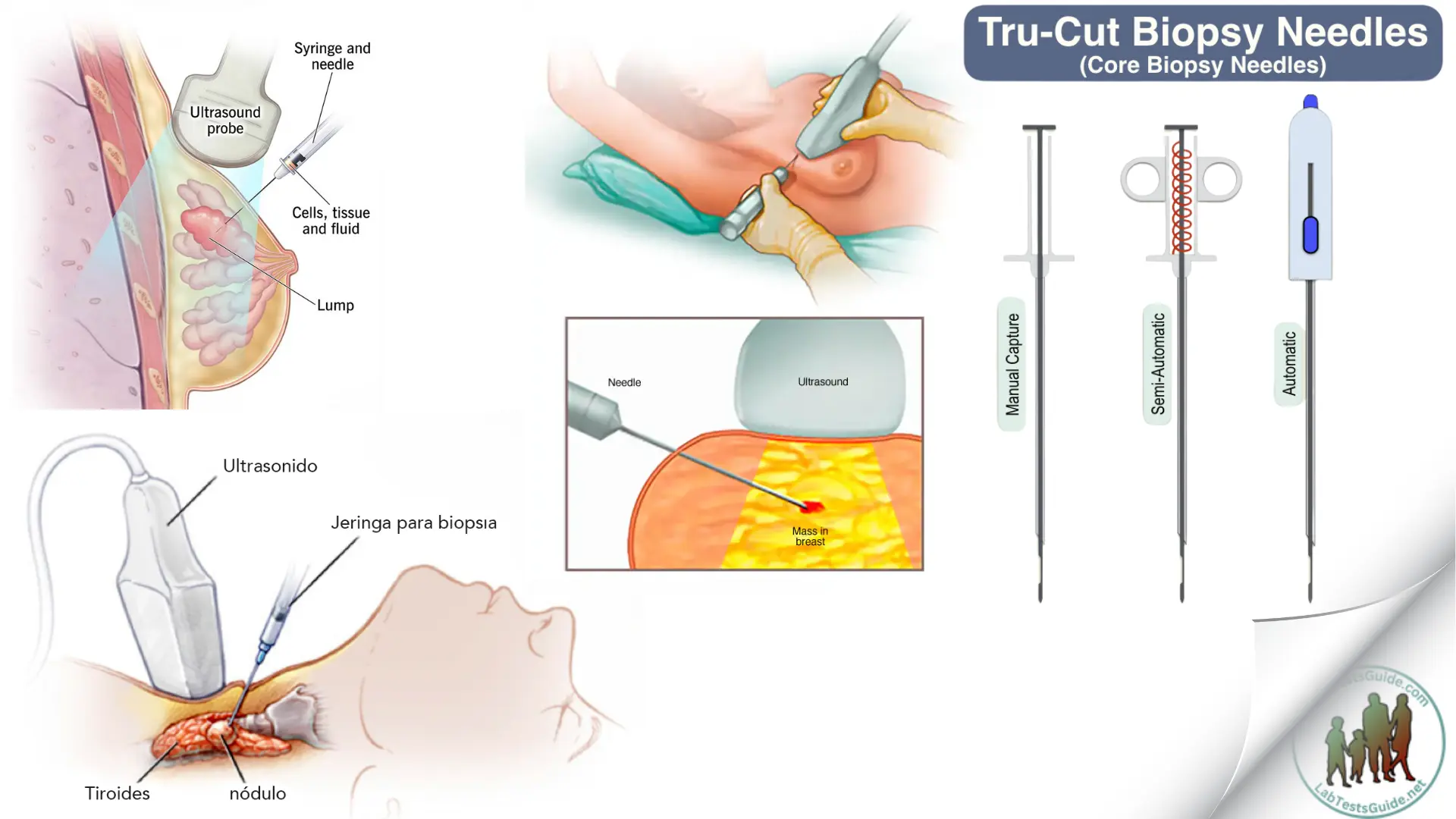
During a needle biopsy, your doctor uses a special needle to remove cells from a suspicious area.

A needle biopsy is often used in tumors that your doctor may feel through your skin, such as suspicious lumps in the breasts and enlarged lymph nodes. When combined with an imaging procedure, such as X-rays, needle biopsy can be used to collect cells from a suspicious area that cannot be felt through the skin.
Needle biopsy procedures include:
- Fine needle aspiration. During fine needle aspiration, a long thin needle is inserted into the suspicious area. A syringe is used to extract fluid and cells for analysis.
- Thick needle biopsy. A larger needle with a cutting tip is used during thick needle biopsy to remove a column of tissue from a suspicious area.
- Vacuum assisted biopsy. During vacuum-assisted biopsy, a suction device increases the amount of fluid and cells that are removed through the needle. This can reduce the number of times the needle must be inserted to collect a suitable sample.
- Image guided biopsy. Image-guided biopsy combines an imaging procedure, such as X-rays, computed tomography (CT), magnetic resonance imaging (MRI) or ultrasound, with a needle biopsy.
The image-guided biopsy allows your doctor to access suspicious areas that cannot be felt through the skin, such as abnormalities in the liver, lungs or prostate. Using real-time images, your doctor can make sure the needle reaches the right place.
You will receive a local anesthetic to numb the biopsy area to minimize pain.
Possible References Used


