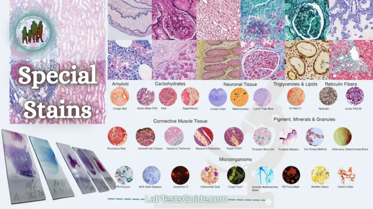
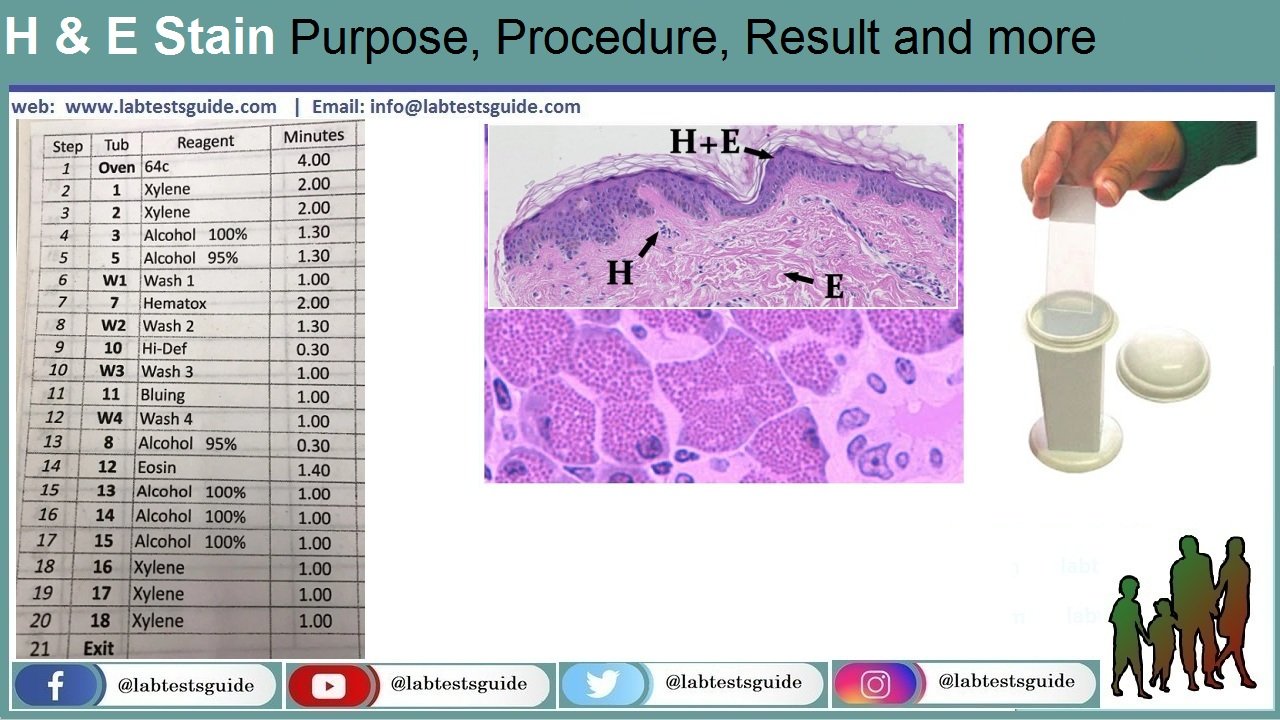
Hematoxylin and eosin staining technique functions to recognize different types of tissues and their morphological changes, especially in cancer diagnosis. Hematoxylin has a deep blue-purple color and stains nucleic acids by a complex, incompletely understood reaction. Eosin is pink and stains proteins nonspecifically. In a typical tissue, nuclei are stained blue, whereas the cytoplasm and extracellular matrix have varying degrees of pink staining.
Principle:
Hematoxylin is extracted from the Hematoxylon campechianum tree on which, by oxidation, hematein is produced from hematoxylin, the dye used in the hematoxylin and eosin staining technique. The addition of a mordant allows the hematein to bind to the anionic elements of the tissues. Hematoxylin without eosin acts as a counterstain in immunohistochemical and hybridization protocols that use colorimetric substrates (such as alkaline phosphatase or peroxidase).
Eosin dye is an acidic dye, therefore it has a negative (eosinophilic) charge. Therefore it stains the basic structures of a cell (acidophilus), giving them a red or pink color, for example, the cytoplasm is positively charged, and therefore it will absorb the eosin dye, and it will appear pink.
Hematoxylin dyes are basic dyes, so they are positively charged. Therefore, it will stain the acidic structures of the tissues and the cellular structures (basophilic), purplish blue. Hematoxylin is not basic in itself. It must be conjugated with a mordant (aluminum salt) before use to strengthen its positive charge and achieve effective binding to tissue components. The mordant, which also defines the color of the stain, will bind to the tissue, then the hematoxylin will bind to the mordant to form a tissue-mordant-hematoxylin complex bond. This will stain the nuclei and chromatin bodies purple.
H&E staining can be classified into three types: progressive, modified progressive, and regressive. Progressive staining takes place without a differentiator to remove any excess dye after adding hematoxylin. This causes background staining to occur on the loaded slides. It is mainly used to dye Mucin. While progressive and regressive Modify, use a differentiator to remove excessive tints. It is used for the demonstration of the kernel content.
Reagents:
- Distilled water
- Alum hematoxylin
- Acid alcohol
- Scott’s tap water
- Eosin dye
Procedure:
The procedure is simplified into several processes: dewaxing, dehydration, hematoxylin, differentiation, bluish, eosin, dehydration, rinsing, coverslip.
- Clean the sections with distilled water.
- Then stain the nuclei with Alum Hematoxylin (from Mayer) to fix the tissue, for about 5 minutes.
- Rinse the stain under mild running water.
- Using the differentiator, 0.3% acid alcohol, and note the end point, that is, the correct end point is after bluish, the background becomes colorless.
- Rinse the stain under running water without problems.
- Rinse the satin in Scott’s tap water substitute, which shortens the time to the correct end point.
- Rinse under running tap water.
- Flood the smear with eosin for 2 minutes, and since eosin is very soluble in water, use a sufficient amount. The over-stained eosin can be removed or washed under running water.
- Dehydrate the smear, rinse, and mount with a clean coverslip.
Results and interpretation:
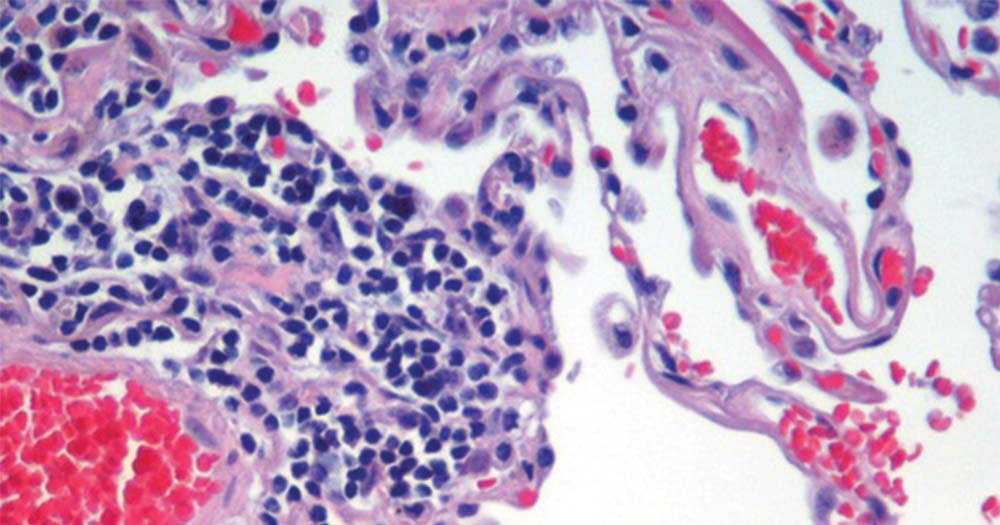
Nuclei stain blue, while cytoplasm and extracellular matrix have varying degrees of pink staining. Well-fixed cells show considerable intranuclear detail. The nuclei show different patterns of heterochromatin condensation (hematoxylin staining) specific to the cell type and the cancer type that are very important from a diagnostic point of view. Nucleoli staining with eosin. If there are abundant polyribosomes, the cytoplasm will have a distinctive blue hue.
Related Articles:
Keywords: haematoxylin and eosin,eosin,hematoxylin and eosin,haematoxylin and eosin staining,haematoxylin and eosin staining protocol,haematoxylin and eosin staining method,haematoxylin and eosin staining technique,hematoxylin and eosin staining procedure,haematoxylin and eosin staining steps,haematoxylin and eosin (h/e) staining,what is haematoxylin and eosin staining,haematoxylin and eosin staining principle,haematoxylin and eosin staining procedure,eosin and hematoxylin,hematoxylin and eosin stain, h and e stain,h and e staining,stain,h and e staining procedure,h&e stain,hematoxylin and eosin staining,haematoxylin and eosin staining,uses of h and e stain,h and e stain purpose,hematoxylin and eosin staining principle,hematoxylin and eosin staining procedure,h and e stain protocol,h and e stain procedure,h and e staining histology,h and e staining principle,procedure of h and e stain,h and e stain preparation,application of h and e stain,hamatoxylin and eosin stain
Possible References Used


