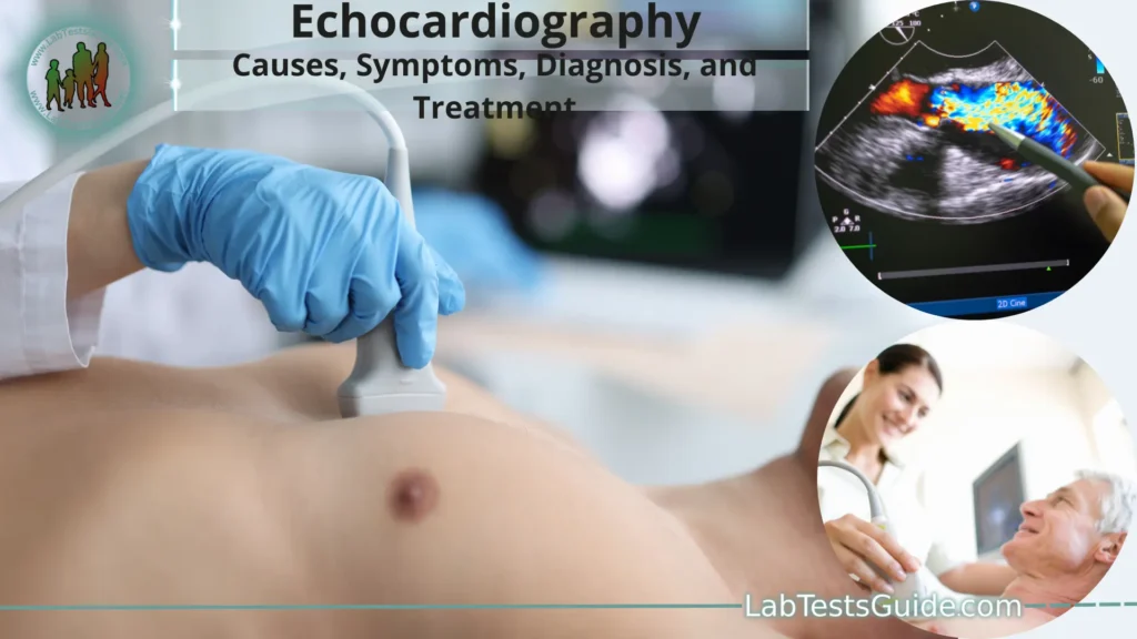
Echocardiography, also known as an echo, is a non-invasive medical imaging technique used to assess the structure and function of the heart. It is an essential tool in cardiology for diagnosing various heart conditions and monitoring heart health. The procedure utilizes high-frequency sound waves (ultrasound) to create real-time images of the heart, its chambers, valves, and blood flow patterns.

What is Echocardiography?
Echocardiography is a medical imaging technique used to visualize the heart’s structures and assess its function. It employs high-frequency sound waves (ultrasound) to create real-time images of the heart, including its chambers, valves, blood vessels, and surrounding tissues. The procedure is safe, non-invasive, and does not involve exposure to ionizing radiation.
During an echocardiogram, a small handheld device called a transducer is placed on the patient’s chest (in the case of a transthoracic echocardiogram) or inserted into the esophagus (in the case of a transesophageal echocardiogram). The transducer emits sound waves that travel into the body and bounce off the heart’s structures. The echoes produced by the bouncing sound waves are captured by the transducer and converted into detailed images by a computer.
Types of Echocardiograms:
The main types of echocardiograms include.
Transthoracic Echocardiogram (TTE):
- This is the most common type of echocardiogram.
- The transducer is placed on the chest’s external surface to obtain images of the heart’s structures through the chest wall.
- It is non-invasive, safe, and painless.
- TTE is used for routine heart evaluations, diagnosing heart conditions, and monitoring heart health.
Transesophageal Echocardiogram (TEE):
- TEE involves inserting the transducer into the esophagus through the mouth.
- This provides closer and clearer images of the heart, particularly the structures that are not well visualized by TTE due to the presence of the lungs or ribs.
- TEE is useful for evaluating heart valves, detecting blood clots, and assessing certain cardiac conditions more accurately.
Stress Echocardiogram:
- Stress echocardiography involves assessing the heart’s function during physical stress (exercise) or with the use of medications that simulate exercise.
- This test helps evaluate the heart’s response to stress, which can unmask certain heart conditions that may not be apparent at rest.
- Stress echocardiography is commonly used for diagnosing coronary artery disease and evaluating overall heart function.
3D Echocardiogram:
- A 3D echocardiogram creates three-dimensional images of the heart, providing more detailed and comprehensive views of the heart’s structures.
- It allows for better visualization of complex cardiac anatomy and can aid in surgical planning and interventions.
Fetal Echocardiogram:
- Fetal echocardiography is performed during pregnancy to assess the heart of the developing fetus.
- It helps detect congenital heart defects and other cardiac abnormalities before birth, allowing for timely management and treatment after delivery.
Contrast Echocardiogram:
- Contrast agents (microbubbles) are used to enhance the visualization of blood flow within the heart.
- This type of echocardiogram is helpful in assessing blood flow patterns and identifying areas of reduced blood supply or abnormalities.
Intraoperative and Transesophageal Echocardiography (TEE):
- Echocardiography is sometimes used during surgery to monitor heart function and guide certain cardiac procedures in real-time.
- TEE is commonly used in cardiac surgery to assist with valve repairs/replacements, congenital heart defect repairs, and other procedures.
Basic Principles of Echocardiography:
Here are the fundamental principles involved in echocardiography.
- Ultrasound Waves: Echocardiography utilizes high-frequency sound waves (ultrasound) beyond the range of human hearing. The ultrasound waves are emitted by a transducer, which is a small handheld device that also receives the echoes produced by the sound waves.
- Sound Wave Reflection: When the ultrasound waves encounter tissue interfaces in the heart, such as between different heart chambers, valves, or blood vessels, some of the sound waves get reflected back (echo) to the transducer.
- Time-of-Flight Measurement: The echocardiography system measures the time taken for the sound waves to travel from the transducer to the tissue interface and back. By knowing the speed of sound in tissues, the system can calculate the distance between the transducer and the reflecting structure.
- Image Formation: The echoes received by the transducer are processed by a computer to create real-time images of the heart. The computer constructs these images based on the timing and strength of the returning echoes. Different tissue densities and interfaces create varying levels of echo intensity, resulting in grayscale images with distinct anatomical details.
- Two-Dimensional Imaging: Standard echocardiography produces two-dimensional (2D) images of the heart, displaying multiple cross-sectional views of the cardiac structures. By moving the transducer to different positions on the chest or using transesophageal echocardiography (TEE), clinicians can obtain various imaging planes and views.
- Doppler Techniques: Echocardiography can also use Doppler techniques to assess blood flow. Doppler ultrasound measures the frequency shift of sound waves reflected off moving red blood cells, allowing evaluation of blood flow direction, velocity, and turbulence. Color Doppler imaging displays blood flow in color, making it easier to visualize blood flow patterns.
- M-Mode (Motion Mode): M-mode is a specialized mode in echocardiography that displays motion of specific structures over time. It provides a single-dimensional view and is useful for precise measurements of cardiac dimensions and motion.
- Echocardiography Artifacts: Echocardiograms may sometimes display artifacts, which are non-cardiac structures or signals that interfere with image quality. Understanding these artifacts is crucial for accurate image interpretation.
- Image Interpretation: Trained healthcare professionals, usually cardiologists or cardiac sonographers, interpret the echocardiographic images. They analyze the structure and function of the heart, assess the valves, measure chamber sizes, evaluate blood flow patterns, and identify any abnormalities or cardiac diseases.
Transthoracic Echocardiography (TTE):
- Preparation: Before the procedure, the patient may be asked to change into a hospital gown to expose the chest. Some electrodes or sticky patches may be attached to the chest to monitor the heart’s electrical activity (ECG) during the examination.
- Patient Positioning: The patient is usually lying down on an examination table on their left side. The sonographer (the trained technician performing the test) applies a special gel to the chest to help transmit the ultrasound waves and eliminate air between the skin and transducer.
- Transducer Placement: The sonographer then places the transducer on different areas of the chest to obtain various views of the heart. The transducer emits ultrasound waves, which penetrate through the chest wall and bounce off the heart’s structures, creating real-time images on a monitor.
- Standard Views: Several standard views are typically obtained during a TTE examination, including the parasternal long-axis, parasternal short-axis, apical four-chamber, and apical two-chamber views. Each view provides specific information about the heart’s anatomy and function.
- M-Mode and Doppler: The sonographer may use M-Mode to obtain precise measurements of cardiac structures and motion. Additionally, Doppler techniques can be applied to assess blood flow, including color Doppler to visualize the direction and velocity of blood flow.
- Image Interpretation: The images produced during TTE are interpreted by a cardiologist or a skilled sonographer. They analyze the heart’s chambers, valves, wall thickness, ejection fraction (a measure of the heart’s pumping function), and assess for any abnormalities or structural heart conditions.
- Diagnostic Uses: TTE is used to diagnose and monitor a wide range of heart conditions, including heart valve abnormalities, heart muscle disorders (cardiomyopathy), congenital heart defects, pericardial diseases (e.g., pericarditis), and heart function assessment in various clinical scenarios.
- Safety and Advantages: TTE is a safe procedure, free from ionizing radiation, and generally well-tolerated by patients. It provides real-time dynamic images, allowing the evaluation of cardiac motion and function.
Transesophageal Echocardiography (TEE):
Here are the key features and aspects of Transesophageal Echocardiography.
- Procedure Preparation: Before the TEE procedure, the patient is usually required to fast for several hours to ensure an empty stomach. In some cases, sedation or anesthesia may be administered to make the procedure more comfortable and to prevent gag reflexes during the insertion of the transducer.
- Transducer Insertion: A specialized transducer (TEE probe) is mounted on a flexible tube and carefully inserted through the mouth, down the esophagus, and into the lower part of the esophagus, closer to the heart. The esophagus lies directly behind the heart, allowing for close proximity to the cardiac structures.
- Imaging Process: The transducer emits ultrasound waves from the esophagus, which bounce off the heart structures and create detailed images on a monitor. The TEE probe is equipped with a transducer at its tip, allowing for high-frequency and high-resolution imaging.
- Detailed Views: TEE provides detailed images of the heart valves, chambers, and great vessels, with excellent visualization of the posterior structures, such as the left atrium and the backside of the heart. This detailed view is particularly beneficial for assessing valvular heart diseases, prosthetic heart valves, and identifying cardiac masses or blood clots.
- Doppler and 3D Imaging: Similar to TTE, TEE can also utilize Doppler techniques to assess blood flow patterns and velocities within the heart. Furthermore, 3D TEE provides volumetric images that offer additional insights into complex cardiac anatomy.
- Real-Time Guidance: TEE is often used in the operating room and during cardiac procedures, providing real-time guidance for interventions such as heart valve repairs or replacements. It allows the medical team to visualize the heart structures in detail and assess the success of the procedure.
- Safety and Precautions: TEE is considered safe, but like any medical procedure, it carries some risks. Potential complications may include sore throat, gag reflex, or rare instances of esophageal injury. These risks are minimized through proper patient preparation, experienced operators, and continuous monitoring during the procedure.
Stress Echocardiography:
The key features and aspects of Stress Echocardiography are as follows.
- Purpose: Stress Echocardiography is primarily used to diagnose or evaluate coronary artery disease (CAD), which is caused by a narrowing or blockage of the coronary arteries that supply blood to the heart muscle. The test helps identify regions of the heart that may not be receiving adequate blood supply during exercise or stress, indicating areas of reduced blood flow (ischemia).
- Stress Induction: Stress is typically induced either through exercise or by administering certain medications that mimic the effects of exercise on the heart. Exercise stress is achieved by having the patient walk on a treadmill or pedal a stationary bicycle while being continuously monitored. Medications like dobutamine or adenosine may be used for pharmacological stress, suitable for patients who cannot exercise.
- Echocardiographic Imaging: During stress, echocardiographic images are obtained before and immediately after the stress-inducing activity. The transducer is placed on the patient’s chest to visualize the heart’s structures and blood flow during different stages of the stress test.
- Comparison of Images: The images acquired before and after stress are compared to identify changes in the heart’s function and blood flow. Normal blood flow is expected to increase during stress, supplying adequate oxygen to the heart muscle. If certain segments of the heart show reduced blood flow (indicating ischemia), it may suggest the presence of CAD.
- Interpretation: A cardiologist or a skilled sonographer interprets the Stress Echocardiography results. The images are analyzed for wall motion abnormalities, which can indicate areas of reduced blood flow or ischemia. The presence and severity of these abnormalities can guide further diagnostic and treatment decisions.
- Uses and Applications: Stress Echocardiography is particularly beneficial for patients with suspected CAD, those with atypical symptoms of heart disease, or those who cannot undergo other stress tests like nuclear stress tests or cardiac catheterization.
- Safety: Stress Echocardiography is considered safe and well-tolerated by most patients. However, it should be performed under the supervision of trained medical professionals, as exercise or certain medications used to induce stress can temporarily increase heart rate and blood pressure.
Advanced Echocardiographic Techniques:
Some of the notable advanced echocardiographic techniques include.
Three-Dimensional Echocardiography (3D Echo):
- 3D echocardiography creates real-time three-dimensional images of the heart, providing a more realistic view of cardiac structures.
- It allows for better visualization of complex anatomies, such as heart valves and congenital heart defects, enhancing diagnostic accuracy.
- 3D Echo aids in surgical planning, guiding interventional procedures, and improving overall image understanding.
Strain Imaging and Speckle Tracking:
- Strain imaging assesses the deformation (strain) of the heart muscle during the cardiac cycle, providing information about regional myocardial function.
- Speckle tracking is a technique used to track unique speckles or markers within the myocardium, enabling quantitative assessment of strain and strain rate.
- Strain imaging helps in early detection of subtle changes in heart function and can be used to monitor cardiotoxic effects of certain medications or conditions.
Contrast Echocardiography:
- Contrast agents (microbubbles) are injected intravenously to enhance the visualization of blood flow within the heart.
- Contrast echocardiography improves the detection of small blood flow abnormalities and assists in identifying areas of reduced blood supply (perfusion defects).
Transesophageal Echocardiography with 3D Imaging (3D TEE):
- Combines the benefits of transesophageal echocardiography (TEE) with real-time three-dimensional imaging.
- 3D TEE provides detailed views of cardiac structures with improved spatial resolution, especially for valve assessments and guiding complex interventions.
Intracardiac Echocardiography (ICE):
- ICE involves placing an echocardiography probe within the heart through a catheter introduced from a blood vessel (usually via the femoral vein).
- It is commonly used during catheter-based procedures, such as atrial septal defect (ASD) closure or left atrial appendage (LAA) occlusion, to guide the interventions in real-time.
2D Speckle Tracking and 3D Strain for Assessing Cardiac Mechanics:
- These advanced techniques allow quantitative assessment of myocardial mechanics, including myocardial strain and strain rate.
- They offer valuable insights into regional and global myocardial function, contributing to a more comprehensive assessment of cardiac performance.
Contrast Harmonic Imaging (CHI):
- CHI is a technique that enhances the contrast between blood and tissue by utilizing harmonic frequencies generated from microbubbles.
- This technique improves endocardial border delineation, facilitating more accurate measurements and assessments.
Pediatric Echocardiography:
Key features and aspects of Pediatric Echocardiography include:
- Age-Appropriate Imaging: Pediatric echocardiography techniques are tailored to the age and size of the child. The imaging protocol varies depending on the child’s age, from neonates to adolescents, to obtain the best possible views of the heart.
- Congenital Heart Defects: Pediatric echocardiography is particularly important for diagnosing and characterizing congenital heart defects, which are structural heart abnormalities present at birth. Echocardiography helps in identifying the type and severity of defects, guiding treatment decisions, and planning for surgical interventions if necessary.
- Growth Adaptations: Pediatric echocardiography takes into account the normal growth and changes in heart anatomy as a child grows. Age-specific reference values are used to assess cardiac dimensions and function, considering the age, height, and weight of the child.
- Fetal Echocardiography: Fetal echocardiography is performed during pregnancy to assess the heart of the developing fetus. It can detect congenital heart defects and other cardiac abnormalities before birth, allowing for early management and planning for specialized care after delivery.
- Non-Invasive and Safe: Pediatric echocardiography is non-invasive, safe, and well-tolerated by children. It does not involve exposure to ionizing radiation, making it ideal for repeated evaluations and long-term follow-up.
- Role in Pediatric Cardiology: Pediatric echocardiography plays a central role in pediatric cardiology by providing comprehensive information about the heart’s structure and function. It aids in the diagnosis of various conditions, including septal defects, valve abnormalities, cardiomyopathies, and other acquired heart diseases.
- Role in Congenital Heart Surgery: Echocardiography is crucial in planning and assessing the outcomes of congenital heart surgeries. It provides detailed information for surgical teams and helps monitor postoperative recovery and potential complications.
- Cardiac Screening: Pediatric echocardiography is often used for cardiac screening in newborns and infants with suspected heart conditions or as part of routine newborn screening programs.
- Pediatric Cardiac Intensive Care: Echocardiography is frequently used in pediatric cardiac intensive care units to monitor critically ill children and assess the effectiveness of treatments.
Echocardiography in Specific Cardiac Conditions:
Here are some specific cardiac conditions where echocardiography is particularly useful.
Valvular Heart Disease:
- Echocardiography is essential for assessing heart valve function and detecting valvular abnormalities such as stenosis (narrowing) or regurgitation (leakage).
- It helps in determining the severity of valve defects, identifying the affected valve(s), and guiding treatment decisions, including the need for valve repair or replacement.
Coronary Artery Disease (CAD):
- Stress Echocardiography is commonly used to evaluate coronary artery disease. It assesses the heart’s response to stress, helping identify areas of reduced blood flow (ischemia) caused by narrowed or blocked coronary arteries.
- Echocardiography can also provide information about wall motion abnormalities and myocardial function in patients with CAD.
Cardiomyopathies:
- Echocardiography aids in the diagnosis and assessment of various types of cardiomyopathies, including dilated cardiomyopathy, hypertrophic cardiomyopathy, and restrictive cardiomyopathy.
- It helps evaluate heart chamber size, wall thickness, and function, providing crucial information for treatment planning and monitoring disease progression.
Congenital Heart Defects:
- Echocardiography is the primary imaging modality for diagnosing congenital heart defects in infants and children. It helps visualize the structural abnormalities in the heart and guides surgical or interventional procedures.
- Fetal echocardiography is used during pregnancy to detect congenital heart defects in the developing fetus.
Heart Failure:
- Echocardiography is essential for assessing heart function and identifying the underlying causes of heart failure.
- It helps evaluate the heart’s pumping capacity (ejection fraction) and provides information about any structural abnormalities or valve dysfunction contributing to heart failure.
Pericardial Diseases:
- Echocardiography is helpful in diagnosing and assessing conditions affecting the pericardium, the lining around the heart.
- It can detect pericardial effusion (accumulation of fluid around the heart) and assess the hemodynamic consequences, such as cardiac tamponade.
Cardiac Masses and Tumors:
- Echocardiography can detect and characterize cardiac masses and tumors, such as myxomas, thrombi (blood clots), or metastatic tumors.
- It helps in determining the location, size, and mobility of the masses and guides decisions regarding surgical excision or further investigations.
Infective Endocarditis:
- Echocardiography plays a critical role in diagnosing infective endocarditis, an infection of the heart valves or inner lining of the heart.
- It can identify vegetations (abnormal growths) on the heart valves and assess the extent of valve damage.
Interventional Echocardiography:
Key aspects of Interventional Echocardiography include.
- Transesophageal Echocardiography (TEE) Guidance: TEE is commonly used during interventional procedures due to its ability to provide clear and detailed images of the heart structures, especially from the esophageal position. TEE provides excellent views of the left atrium, left ventricle, and the posterior heart structures, making it particularly useful in guiding interventions involving these areas.
- Transcranial Doppler (TCD) Monitoring: In certain procedures, TCD may be used to monitor cerebral blood flow during interventional procedures that may involve manipulation of the aorta or other structures near the brain.
- Structural Heart Interventions: Interventional Echocardiography is extensively used in structural heart interventions, such as transcatheter aortic valve replacement (TAVR), transcatheter mitral valve repair (MitraClip), patent foramen ovale (PFO) closure, atrial septal defect (ASD) closure, and left atrial appendage (LAA) occlusion. Real-time TEE imaging helps in proper device positioning and assessment of device function.
- Intracardiac Procedures: In certain cases, echocardiography may be used to guide intracardiac procedures, such as biopsy of cardiac masses, closure of ventricular septal defects, or evaluation of complications related to pacemaker or defibrillator placement.
- Percutaneous Coronary Interventions (PCI): Echocardiography can be employed to assess left ventricular function during PCI, particularly in high-risk cases or when there is concern about coronary blood flow and myocardial viability.
- Electrophysiology Procedures: During certain electrophysiology studies and catheter ablation procedures, echocardiography may be used to visualize cardiac anatomy, identify anatomical landmarks, and monitor cardiac function.
- Real-Time Monitoring: Interventional Echocardiography allows the interventionalist to monitor changes in cardiac anatomy and function in real-time during the procedure. It aids in identifying potential complications and making necessary adjustments.
- Assessment of Complications: In the event of complications during an interventional procedure, echocardiography can be used to evaluate and diagnose issues like valve leaks, pericardial effusion, or device malposition.
Artifacts and Pitfalls in Echocardiography:
Some common artifacts and pitfalls in echocardiography include.
- Reverberation Artifact: This occurs when ultrasound waves bounce back and forth between two strong reflectors, creating multiple echoes on the image. It can cause false echoes and distort the image.
- Shadowing Artifact: Shadowing occurs when ultrasound waves are blocked by dense structures, such as bone or calcifications, leading to a lack of information beyond these structures.
- Attenuation Artifact: Attenuation is the reduction of ultrasound signal intensity as it passes through tissues. Significant attenuation can lead to weakened signals and difficulty visualizing structures deeper within the body.
- Side Lobe Artifact: Side lobes are secondary sound beams that may create false echoes, particularly when imaging near metal objects, such as prosthetic heart valves.
- Mirror Artifact: This artifact can occur in transesophageal echocardiography when strong reflectors, such as the aortic valve, create a false image on the opposite side of the true structure.
- Edge Artifact (Refraction Artifact): Edge artifacts occur when the ultrasound beam encounters sharp interfaces, causing deflection and distortion of the beam, resulting in inaccurate image representation.
- Motion Artifact: Patient or probe motion during image acquisition can lead to blurring and loss of image clarity.
- Respiratory Artifact: Breathing or respiratory motion can cause image distortion, especially in transthoracic echocardiography.
- Gain Setting Errors: Incorrect gain settings can lead to over-amplification or under-amplification of echoes, affecting image brightness and contrast.
- Reverberation Between Catheter Tip and Valve: In intracardiac echocardiography (ICE) during catheter-based procedures, echoes can be generated between the catheter tip and heart valve, potentially obscuring valve visualization.
- Electrical Interference: Electrical noise from other devices or environmental sources can interfere with the ultrasound signals, affecting image quality.
- Patient-Specific Factors: Patient anatomy, body habitus, and lung tissue can affect acoustic windows and the ability to obtain clear images.
- Instrument Settings: Improper instrument settings, such as depth, focus, or transducer frequency, can lead to suboptimal image quality.
Echocardiography in Emergency and Critical Care Settings:
Some of the key uses of echocardiography in emergency and critical care settings include:
- Rapid Diagnosis of Cardiac Conditions: Echocardiography can quickly assess cardiac function and identify conditions such as acute myocardial infarction (heart attack), pericardial effusion (fluid around the heart), cardiac tamponade (compression of the heart by fluid), and acute heart failure.
- Evaluation of Shock: In critically ill patients with shock, echocardiography helps determine the underlying cause, such as cardiogenic shock (heart pump failure) or distributive shock (septic shock, anaphylactic shock).
- Assessment of Cardiac Function: Echocardiography provides information on left ventricular function, ejection fraction, and wall motion abnormalities, which are crucial in evaluating heart function and guiding therapy.
- Hemodynamic Monitoring: Echocardiography allows for the assessment of cardiac output, intracardiac pressures, and valvular function, assisting in the management of patients requiring hemodynamic support.
- Evaluation of Cardiac Arrest: In cases of cardiac arrest, echocardiography can be used during cardiopulmonary resuscitation (CPR) to assess cardiac activity, identify reversible causes of arrest, and guide resuscitative efforts.
- Trauma Evaluation: Echocardiography aids in evaluating cardiac injuries and pericardial effusions in trauma patients, helping in rapid decision-making and intervention.
- Guiding Procedures: Echocardiography can be used to guide central venous catheter placement, pericardiocentesis (fluid drainage from the pericardial sac), and other bedside procedures.
- Assessment of Fluid Status: Echocardiography assists in evaluating intravascular volume and fluid responsiveness in critically ill patients.
- Intraoperative Monitoring: In cardiac surgery or other high-risk surgeries, intraoperative echocardiography helps assess heart function and guide surgical decisions.
- Monitoring Response to Treatment: Serial echocardiography can be used to monitor the response to treatment and guide adjustments in critical care settings.
- Emergency Transport: Portable or handheld echocardiography devices (e.g., point-of-care ultrasound) enable rapid assessment during patient transport or in resource-limited settings.
FAQs:
What is echocardiography?
Echocardiography is a non-invasive medical imaging technique that uses ultrasound waves to create real-time images of the heart’s structures and assess its function. It is a valuable tool in cardiology for diagnosing and managing various heart conditions.
How is echocardiography performed?
Echocardiography is performed by placing a transducer on the patient’s chest (transthoracic echocardiography) or inserting it into the esophagus (transesophageal echocardiography). The transducer emits ultrasound waves that bounce off the heart structures, creating images on a monitor.
What are the different types of echocardiograms?
The main types of echocardiograms include transthoracic echocardiogram (TTE), transesophageal echocardiogram (TEE), stress echocardiogram, fetal echocardiogram, and contrast echocardiogram, among others.
What are the advantages of echocardiography?
Echocardiography is safe, non-invasive, and does not involve exposure to ionizing radiation. It provides real-time dynamic imaging of the heart, allowing for the assessment of cardiac function and structures.
How is echocardiography used in diagnosing heart conditions?
Echocardiography is used to diagnose various heart conditions, including valvular heart disease, coronary artery disease, cardiomyopathies, congenital heart defects, and pericardial diseases. It provides valuable information about the heart’s structure and function.
Is echocardiography safe?
Yes, echocardiography is considered safe and well-tolerated by most patients. It does not involve radiation exposure or significant risks when performed by trained professionals.
How does stress echocardiography work?
Stress echocardiography involves evaluating the heart’s response to stress induced by exercise or medications. It helps diagnose coronary artery disease and assesses areas of reduced blood flow (ischemia) during stress.
What is the role of echocardiography in pediatric cardiology?
Echocardiography is crucial in pediatric cardiology for diagnosing congenital heart defects, assessing heart function in children, and guiding treatment decisions. It is tailored to the age and size of the child.
How is echocardiography used in emergency and critical care settings?
Echocardiography is used in emergency and critical care settings for rapid diagnosis of cardiac conditions, assessment of shock, evaluation of cardiac function, and guiding interventions and resuscitative efforts.
Can echocardiography guide cardiac procedures?
Yes, echocardiography can guide cardiac procedures, including structural heart interventions (e.g., TAVR, MitraClip), electrophysiology procedures, and intracardiac procedures. Real-time imaging helps in device positioning and assessing function during the procedure.
Is echocardiography affected by artifacts?
Yes, echocardiography may be affected by artifacts, which can impact image quality and interpretation. Common artifacts include reverberation, shadowing, and motion artifacts.
How does echocardiography contribute to cardiac surgery?
Echocardiography plays a significant role in cardiac surgery by providing detailed preoperative assessments, intraoperative guidance, and postoperative monitoring of surgical outcomes.
Is echocardiography used during pregnancy?
Yes, fetal echocardiography is used during pregnancy to assess the heart of the developing fetus and detect congenital heart defects and other cardiac abnormalities before birth.
What is the future of echocardiography?
The future of echocardiography is likely to involve further advancements in imaging technology, including improved 3D and strain imaging, as well as greater integration with artificial intelligence for automated analysis and decision support.
Conclusion:
In conclusion, echocardiography is a powerful and versatile imaging modality used in cardiology for assessing the heart’s structure and function. It offers real-time, non-invasive, and safe visualization of cardiac anatomy, aiding in the diagnosis and management of a wide range of heart conditions, from congenital heart defects and valvular abnormalities to coronary artery disease and cardiomyopathies. Echocardiography’s role extends across various clinical settings, from routine screenings to emergency and critical care situations, guiding interventional procedures, surgical planning, and providing valuable information for patient care. As technology continues to advance, echocardiography is poised to further enhance cardiac imaging capabilities and contribute to improved patient outcomes in the future.
Possible References Used


