A CT scan, or computed tomography scan, is a medical imaging technique that uses X-rays and computer processing to create detailed cross-sectional images of the inside of the body. It is a valuable diagnostic tool used by healthcare professionals to visualize various structures and tissues within the body, including the brain, chest, abdomen, pelvis, and extremities. CT scans provide a more detailed and comprehensive view of the body than traditional X-rays.

Key Points:
- CT stands for Computed Tomography, a medical imaging technique.
- CT scans use X-rays to create detailed cross-sectional images of the body.
- They offer a more detailed view than conventional X-rays.
- CT scans are used for diagnosing and monitoring various medical conditions.
- The process involves X-ray generation, detection, and computer reconstruction.
- CT scans can create 3D representations of the body’s internal structures.
- Different types of CT scans focus on specific areas like the head, chest, or abdomen.
- CT angiography is used to visualize blood vessels.
- Musculoskeletal CT scans focus on bones and joints.
- CT scans can identify and stage cancerous tumors.
- They are valuable in trauma situations for assessing injuries.
- CT scans are crucial for detecting and evaluating strokes.
- They help diagnose pulmonary conditions like pneumonia.
- Abdominal CT scans assist in diagnosing diseases of the digestive system.
- Contrast agents are sometimes used to enhance the visibility of certain tissues.
- CT scans have a small radiation exposure, but safety measures minimize risk.
- Special considerations apply to pregnant women and children undergoing CT scans.
- Patients may need to fast or follow specific instructions before the procedure.
- During the scan, patients lie on a table that moves through the CT machine.
- The duration of a CT scan varies but is generally relatively quick.
- Radiologists interpret CT scan results to diagnose medical conditions.
- Normal vs. abnormal findings are identified in the images.
- Radiologists report their findings to referring healthcare providers.
- CT technology continually advances, with improved image quality and reduced radiation.
- CT scans play a vital role in modern healthcare, aiding in diagnosis, treatment planning, and monitoring patients’ progress.
What Is a CT Scan :
A CT scan, or Computed Tomography scan, is a medical imaging technique that uses X-rays and computers to create detailed cross-sectional images of the body’s internal structures, providing valuable diagnostic information for various medical conditions.
History of CT Scanning:
- 1967: The Invention – The first CT scanner, called the “EMI Scanner,” was invented by Sir Godfrey Hounsfield at EMI Laboratories in England.
- 1971: First Clinical Scan – The first clinical CT scan was performed on a patient’s head, revolutionizing medical imaging.
- 1974: Nobel Prize – Godfrey Hounsfield and Allan Cormack were awarded the Nobel Prize in Physiology or Medicine for their work on the development of CT scanning.
- 1970s: Early Clinical Use – CT scanning quickly gained popularity in hospitals for diagnosing brain and head injuries.
- 1980s: Advancements – CT technology improved, with the introduction of helical (spiral) CT and higher-resolution images.
- 1990s: Expanding Applications – CT scans became more versatile, used for diagnosing various conditions, including heart disease and cancer.
- 1998: Multislice CT – Siemens introduced the first multislice CT scanner, allowing faster and more detailed imaging.
- 2000s: Reduced Radiation – Advances in technology led to dose reduction techniques, minimizing radiation exposure to patients.
- 2010s: Dual-Energy CT – Dual-energy CT scanners emerged, providing enhanced tissue differentiation.
- 2013: Photon-Counting CT – Experimental photon-counting CT scanners were developed, potentially further reducing radiation exposure.
- Present Day: AI Integration – Artificial intelligence and machine learning are increasingly used to assist in image analysis and interpretation.
- Future Prospects: Ongoing Research – Ongoing research aims to improve CT technology further, including faster scans, reduced radiation, and enhanced diagnostic capabilities.
Purpose and Significance of CT Scans:
The purpose and significance of CT (Computed Tomography) scans in healthcare are extensive, and they play a crucial role in various medical scenarios. Here’s an overview of their purpose and significance:
Purpose:
- Diagnosis: CT scans are primarily used for diagnosing a wide range of medical conditions. They provide detailed cross-sectional images of the body, allowing healthcare professionals to detect abnormalities, injuries, and diseases.
- Staging: In cancer care, CT scans help determine the extent and stage of tumors, aiding in treatment planning and prognosis assessment.
- Monitoring: Patients with chronic conditions or diseases can be monitored over time using CT scans to assess the progression or response to treatment.
- Emergency Medicine: In emergency situations, CT scans quickly assess injuries, such as head trauma, internal bleeding, fractures, and spinal cord injuries, enabling rapid medical decisions.
- Guiding Procedures: CT-guided procedures, like biopsies or drainage of abscesses, ensure accurate targeting of specific tissues or areas within the body.
- Vascular Imaging: CT angiography visualizes blood vessels, helping diagnose conditions such as aneurysms, stenosis, and vascular diseases.
- Bone and Joint Assessment: Musculoskeletal CT scans are valuable for evaluating bone fractures, joint injuries, and orthopedic conditions.
Significance:
- Early Detection: CT scans can identify diseases and abnormalities at an early stage, allowing for timely intervention and better outcomes.
- Precision: They provide highly detailed and accurate images, aiding in precise diagnosis and treatment planning.
- Minimally Invasive: CT-guided procedures are minimally invasive, reducing the need for open surgeries and associated risks.
- Patient-Centric: CT scans are non-invasive, less uncomfortable than some other diagnostic procedures, and are often preferred by patients.
- Treatment Planning: Surgeons and oncologists rely on CT scans to plan surgeries and radiation therapy, ensuring precise targeting of affected areas.
- Follow-up Monitoring: CT scans enable continuous monitoring of treatment effectiveness and disease progression.
- Research and Development: CT technology advancements, including reduced radiation doses and faster scanning, contribute to ongoing medical research and innovation.
- Emergency Care: In emergency medicine, CT scans can quickly assess injuries, enabling rapid and life-saving interventions.
- Improved Outcomes: By providing valuable diagnostic information, CT scans contribute to improved patient outcomes and quality of care.
How CT Scans Work:
The principles of a CT (Computed Tomography) scan involve the use of X-rays and computer technology to create detailed cross-sectional images of the body’s internal structures. Here are the key principles of how CT scans work:
- X-ray Generation: CT scans start with the generation of X-rays. X-rays are a form of electromagnetic radiation.
- X-ray Source: In a CT scanner, there’s an X-ray source that emits a narrow, fan-shaped beam of X-rays.
- X-ray Detection: On the opposite side of the scanner, there’s an array of X-ray detectors. These detectors measure the intensity of X-rays that pass through the body.
- Multiple Angles: The patient lies on a table that can move into and out of the scanner. As the patient moves through the machine, the X-ray source and detectors rotate around the body. This process allows for X-ray images to be taken from multiple angles around the body.
- Data Collection: As the X-ray source and detectors rotate, they collect a series of X-ray images or “slices.” These slices are thin cross-sections of the body.
- Computer Reconstruction: The collected X-ray data is sent to a computer, which uses complex mathematical algorithms to reconstruct the individual slices into a 3D image of the body.
- Cross-Sectional Images: The reconstructed images are typically displayed as cross-sectional slices, each representing a different depth within the body. These images provide detailed views of organs, tissues, and structures.
- Density Differences: CT scans work based on the principle that different tissues in the body absorb X-rays to varying degrees. Dense tissues like bones absorb more X-rays and appear white on the images, while less dense tissues like muscles and organs absorb fewer X-rays and appear darker.
- Contrast Agents: In some cases, contrast agents (often iodine-based) are used to enhance the visibility of certain structures or blood vessels. These contrast agents are either ingested, injected intravenously, or introduced through other means, and they highlight specific areas on the CT scan.
- 3D Visualization: The reconstructed images can be used to create 3D representations of the scanned area, providing a more comprehensive view of the anatomy.
Types of CT Scans:
CT (Computed Tomography) scans come in various types, each designed to focus on specific areas of the body or provide specialized information. Here are some common types of CT scans:
- Head CT (Cranial CT): This type of CT scan focuses on the head and brain. It is used to diagnose and assess conditions such as head injuries, strokes, brain tumors, and bleeding within the brain.
- Chest CT (Thoracic CT): A chest CT scan examines the chest area, including the lungs, heart, blood vessels, and surrounding structures. It is used to detect and evaluate conditions like lung cancer, pulmonary embolism, and lung infections.
- Abdomen and Pelvis CT: This scan encompasses the abdominal and pelvic regions, including the liver, kidneys, spleen, pancreas, and reproductive organs. It is helpful for diagnosing abdominal pain, evaluating abdominal trauma, and detecting tumors or abnormalities.
- CT Angiography (CTA): CTA is used to visualize blood vessels throughout the body, including those in the brain, neck, chest, abdomen, and extremities. It aids in diagnosing vascular conditions like aneurysms, stenosis, and blood clots.
- Musculoskeletal CT: Musculoskeletal CT scans focus on the bones and joints, making them valuable for assessing fractures, joint injuries, bone infections, and musculoskeletal disorders.
- Cardiac CT (Coronary CT Angiography – CTA): Cardiac CT is specialized for imaging the heart and coronary arteries. It helps in diagnosing coronary artery disease, heart valve issues, and congenital heart defects.
- Virtual Colonoscopy (CT Colonography): This scan is a non-invasive alternative to traditional colonoscopy. It’s used to screen for colorectal cancer and assess the colon for polyps or other abnormalities.
- Spinal CT: Spinal CT scans focus on the spine and spinal cord. They are used to evaluate spinal injuries, herniated discs, spinal tumors, and other spinal conditions.
- Sinus CT (Sinusitis CT): This type of CT scan examines the sinuses in the head and is used to diagnose sinusitis, nasal polyps, and other sinus-related issues.
- Temporal Bone CT: Temporal bone CT scans assess the structures of the ear and are commonly used to diagnose conditions affecting the ear, such as hearing loss, infections, and tumors.
- Pulmonary CT: This type of CT scan is used to evaluate the lungs in detail, helping in the diagnosis and monitoring of lung diseases, nodules, and lung cancer.
- Adrenal Gland CT: Adrenal CT scans focus on the adrenal glands located on top of the kidneys. They are used to assess adrenal tumors, hormone-related disorders, and other adrenal gland abnormalities.
CT Scan Preparation and Safety:
CT (Computed Tomography) scan preparation and safety measures are essential to ensure a successful and safe imaging procedure.
Patient Preparation:
- Follow Instructions: Patients should carefully follow any preparation instructions provided by their healthcare provider or the imaging facility. Instructions may include fasting, dietary restrictions, or discontinuation of specific medications.
- Contrast Agents: If a contrast agent is required for the scan, patients should inform their healthcare provider of any allergies, particularly iodine allergies, and any previous reactions to contrast agents.
- Medical History: Patients should inform the radiology technologist and healthcare provider about their medical history, including any underlying health conditions, recent surgeries, or pregnancy.
- Pregnancy: Pregnant women should inform their healthcare provider and the radiology department, as the use of ionizing radiation in CT scans may pose risks to the developing fetus. In some cases, alternative imaging methods may be considered.
- Metal Objects: Patients should remove all metal objects and jewelry before the scan, as these can interfere with the imaging process.
- Clothing: Patients may be asked to wear a hospital gown or clothing without metal zippers or buttons to avoid interference with the scan.
- Contrast Injection: If a contrast agent is needed, it is typically administered intravenously (IV) during the scan. Patients may experience a warm sensation or metallic taste when the contrast is injected.
Radiation Safety:
- Radiation Dose: CT scans involve ionizing radiation, so the radiation dose should be kept as low as reasonably achievable (ALARA). Advanced CT scanners use dose reduction techniques to minimize radiation exposure.
- Pediatric Considerations: Special attention is given to pediatric patients to ensure appropriate radiation doses, as children are more sensitive to radiation.
- Shielding: Lead aprons and shields may be used to protect parts of the body not being imaged from unnecessary radiation exposure.
- Allergies: The contrast agent used in CT scans can cause allergic reactions in some individuals. Medical staff should be prepared to manage potential allergic responses.
- Monitoring: During the scan, a radiology technologist or nurse typically monitors the patient and can communicate with them through an intercom system.
- Intravenous Lines: If a contrast agent is used, an intravenous (IV) line will be inserted into a vein before the scan. This is typically done by a trained healthcare professional.
- Post-Scan Care: After the scan, patients may be monitored for any delayed reactions or side effects from the contrast agent.
- Alternative Scans: In certain cases, when the risks of radiation exposure outweigh the benefits, alternative imaging methods like MRI or ultrasound may be recommended.
Contrast Media and Allergies:
- Types of Contrast Media: There are two main types of contrast media: iodinated contrast for CT scans and gadolinium-based contrast for MRI scans.
- Allergic Reactions: Allergic reactions to contrast media are relatively rare but can range from mild to severe. Common reactions include itching, rash, and hives.
- Anaphylactic Reactions: In rare cases, severe allergic reactions known as anaphylaxis can occur. These reactions can be life-threatening and may cause difficulty breathing, a drop in blood pressure, or loss of consciousness.
- Pre-existing Allergies: Individuals with a history of allergies, especially to iodine-based substances, are at a slightly higher risk of having an allergic reaction to contrast media.
- Kidney Function: Patients with impaired kidney function may be at risk for a condition called contrast-induced nephropathy when using iodinated contrast. This is more of a concern for individuals with pre-existing kidney problems.
- Risk Assessment: Before administering contrast media, healthcare providers typically assess a patient’s medical history, including any prior contrast reactions and kidney function, to determine the safest approach.
- Preventive Measures: In some cases, healthcare providers may prescribe medications such as antihistamines or corticosteroids to reduce the risk of allergic reactions.
- Informed Consent: Patients should be informed about the potential risks and benefits of using contrast media and may be asked to sign an informed consent form before the procedure.
- Communication: Patients should communicate any allergies or concerns about contrast media to their healthcare provider. This information is crucial for ensuring patient safety.
- Monitoring: During and after the imaging procedure, patients are typically monitored for any signs of allergic reactions or adverse effects. Medical staff should be prepared to respond to emergencies.
Applications of CT Scans:
CT (Computed Tomography) scans have a wide range of applications in healthcare, making them one of the most versatile and valuable imaging tools. Here are some of the primary applications of CT scans:
- Diagnosis and Staging of Cancer: CT scans are essential for detecting and diagnosing various types of cancer, including lung, liver, kidney, and abdominal cancers. They also help in staging, determining the extent of cancer, and assessing the response to treatment.
- Head and Brain Imaging: CT scans of the head are used to diagnose conditions such as head injuries, strokes, brain tumors, bleeding in the brain, and other neurological disorders.
- Chest Imaging: CT scans of the chest are employed to evaluate lung conditions such as lung cancer, pulmonary embolism, pneumonia, and interstitial lung disease.
- Abdominal and Pelvic Imaging: These scans are instrumental in diagnosing and evaluating conditions of the abdominal and pelvic regions, including gastrointestinal issues, kidney stones, liver diseases, and reproductive system disorders.
- Vascular Imaging (CT Angiography): CT angiography is used to visualize blood vessels and diagnose conditions such as aneurysms, stenosis, and blood clots. It is especially valuable in assessing coronary arteries.
- Musculoskeletal Imaging: CT scans are used to assess bone fractures, joint injuries, orthopedic conditions, and musculoskeletal disorders.
- Cardiac Imaging: Cardiac CT scans, specifically coronary CT angiography (CTA), are employed to assess coronary artery disease, congenital heart defects, and other heart conditions.
- Virtual Colonoscopy (CT Colonography): This minimally invasive alternative to traditional colonoscopy is used to screen for colorectal cancer and assess the colon for polyps or other abnormalities.
- Spinal Imaging: CT scans of the spine help diagnose spinal injuries, herniated discs, spinal tumors, and other spinal conditions.
- Sinus Imaging: Sinus CT scans are used to diagnose sinusitis, nasal polyps, and other sinus-related issues.
- Trauma Assessment: In emergency medicine, CT scans are crucial for rapidly evaluating traumatic injuries, including head injuries, fractures, internal bleeding, and chest trauma.
- Guiding Medical Procedures: CT-guided procedures, such as biopsies, drainage of abscesses, and tumor ablations, provide precise targeting of tissues or lesions.
- Preoperative Planning: Surgeons use CT scans for preoperative planning in various surgeries, ensuring accurate and efficient procedures.
- Pediatric Imaging: CT scans are used in pediatric care to diagnose and monitor various medical conditions in children while taking into consideration radiation dose reduction techniques.
- Research and Development: CT technology continues to advance, contributing to medical research and the development of new diagnostic techniques.
Procedures of CT Scans:
The procedures involved in a CT (Computed Tomography) scan include several key steps, including patient positioning, the scanning process, duration, comfort, and more. Here’s an overview of the typical procedure:
1. Scheduling and Preparation:
- Patients receive instructions regarding any specific preparations, such as fasting or discontinuing certain medications, from their healthcare provider or the imaging facility.
- Patients are informed about the use of contrast agents and potential allergies or reactions.
- If necessary, patients may change into a hospital gown and remove metal objects and jewelry before the scan.
2. Patient Positioning:
- Patients are positioned on a motorized table that can move in and out of the CT scanner.
- Proper positioning is essential to ensure that the area of interest is within the scanner’s field of view.
3. Contrast Agent (if required):
- If a contrast agent is needed for the scan, it may be administered intravenously (IV), orally, or rectally, depending on the type of scan and the area being examined.
- Intravenous contrast is the most common and is usually administered through an IV line inserted into a vein.
4. Scanning Process:
- The CT scanner consists of an X-ray source and an array of detectors.
- The scanner rotates around the patient, capturing multiple X-ray images from different angles.
- These images, or “slices,” are thin cross-sections of the body.
5. Duration of the Scan:
- The duration of a CT scan varies depending on the type of scan, the area being examined, and the complexity of the procedure.
- Most CT scans are relatively quick, lasting anywhere from a few seconds to a few minutes.
6. Monitoring:
- During the scan, a radiology technologist or nurse typically monitors the patient from an adjacent control room.
- Patients may be able to communicate with the staff through an intercom system.
7. Contrast Injection (if required):
- If contrast is used, it is typically injected into the patient’s vein during the scan. Patients may experience a warm sensation or metallic taste during the injection.
8. Breath-Holding and Instructions:
- Patients may be instructed to hold their breath briefly during certain parts of the scan to minimize motion artifacts and ensure clear images.
- Technologists may provide instructions on specific body positions or movements as needed.
9. Comfort and Safety:
- Patient comfort is a priority, and modern CT scanners are designed to minimize discomfort.
- Safety measures, including shielding, are used to protect parts of the body from unnecessary radiation exposure.
10. Post-Scan Monitoring:
- After the scan, patients are typically monitored for any immediate reactions or side effects, especially if contrast was used.
11. Image Reconstruction:
- The collected X-ray data is sent to a computer, which reconstructs the individual slices into 3D images that can be viewed and analyzed by a radiologist.
12. Interpretation and Reporting:
- A radiologist interprets the images, looking for abnormalities and making a diagnosis.
- A report is generated and sent to the referring healthcare provider.
Interpreting CT Scan Results:
Interpreting CT (Computed Tomography) scan results requires specialized training and expertise typically provided by radiologists or other medical professionals with experience in medical imaging. Here’s an overview of the process of interpreting CT scan results:
- Image Review: After the CT scan is performed, a series of cross-sectional images or “slices” is generated by the computer. These images are typically displayed on a computer monitor.
- Image Navigation: Radiologists use specialized software to navigate through the images, allowing them to view different slices and sections of the body. They can adjust the orientation and contrast of the images to gain a better understanding of the anatomy.
- Identification of Structures: Radiologists systematically identify and evaluate various anatomical structures, including bones, organs, blood vessels, and soft tissues. They assess the density and characteristics of these structures.
- Comparison: In some cases, prior CT scans or imaging studies may be available for comparison. This helps radiologists assess changes over time, such as tumor growth or the progression of a disease.
- Abnormalities and Findings: Radiologists look for any abnormalities, anomalies, or pathological conditions. These may include tumors, lesions, fractures, inflammation, fluid collections, or other issues.
- Quantitative Analysis: In addition to visual assessment, radiologists may perform quantitative measurements, such as measuring the size of a tumor or the degree of stenosis in blood vessels.
- Clinical Context: Radiologists consider the clinical context provided by the referring healthcare provider. Patient history, symptoms, and specific clinical questions guide the interpretation process.
- Documentation: Radiologists document their findings in a formal radiology report. This report includes detailed descriptions of any abnormalities, their location, size, and characteristics.
- Recommendations: Based on their interpretation, radiologists may provide recommendations for further diagnostic tests or procedures, such as additional imaging studies, biopsies, or consultations with specialists.
- Communication: Radiology reports are typically sent to the referring healthcare provider, who then discusses the findings and recommendations with the patient. The healthcare provider uses the CT scan results to make treatment decisions.
- Timeliness: In urgent or emergency cases, radiologists may provide preliminary findings quickly to assist in immediate patient care. A more detailed report is often generated later.
- Continuous Learning: Radiologists undergo ongoing training and peer review to ensure accuracy and maintain their skills in interpreting CT scans.
Radiologist’s Role:
A radiologist plays a pivotal role in the interpretation of medical images, including CT scans. Their specialized training and expertise make them essential members of the healthcare team. Here’s an overview of the radiologist’s role in the context of CT scans:
- Image Interpretation: Radiologists are responsible for reviewing and interpreting the CT scan images. They meticulously assess the cross-sectional images to identify any abnormalities, anomalies, or pathological conditions.
- Anatomy Assessment: Radiologists analyze the anatomical structures within the images, including bones, organs, blood vessels, and soft tissues. They evaluate the density, size, shape, and characteristics of these structures.
- Abnormality Detection: Radiologists look for signs of disease, injury, or other medical conditions. These may include tumors, lesions, fractures, inflammation, fluid collections, or other abnormalities.
- Quantitative Analysis: Radiologists may perform quantitative measurements when necessary. For example, they may measure the size of a tumor, assess the degree of stenosis in blood vessels, or calculate other quantitative parameters to aid in diagnosis and treatment planning.
- Clinical Context: Radiologists consider the clinical context provided by the referring healthcare provider. This includes the patient’s medical history, symptoms, and specific clinical questions that guide the interpretation process.
- Comparison to Previous Imaging: In cases where prior CT scans or other imaging studies are available, radiologists compare the current images to the previous ones. This helps them assess changes over time, such as tumor growth or the progression of a disease.
- Documentation: Radiologists document their findings in a formal radiology report. This report includes detailed descriptions of any abnormalities, their location, size, and characteristics.
- Recommendations: Based on their interpretation, radiologists may provide recommendations for further diagnostic tests or procedures. These recommendations assist referring healthcare providers in making treatment decisions.
- Communication: Radiologists collaborate with referring healthcare providers, such as primary care physicians, specialists, and surgeons, to discuss imaging findings and recommendations. Effective communication is vital for coordinating patient care.
- Continuous Learning: Radiologists engage in ongoing education, training, and peer review to ensure their skills and knowledge remain up-to-date. Advances in imaging technology and medical knowledge require radiologists to continuously expand their expertise.
- Subspecialization: Some radiologists choose to subspecialize in specific areas, such as neuroradiology, musculoskeletal radiology, or interventional radiology. Subspecialists focus on particular types of imaging and related procedures.
- Quality Control: Radiologists are involved in quality control and quality assurance efforts within imaging departments to maintain high standards of image quality and patient safety.
Normal vs. Abnormal Findings:
In the context of CT scans and medical imaging, radiologists are trained to distinguish between normal and abnormal findings. Here’s a breakdown of how they differentiate between the two:
Normal Findings:
- Expected Anatomy: Radiologists first identify and evaluate the expected anatomical structures within the CT scan images. These include bones, organs, blood vessels, and soft tissues.
- Consistency with Known Anatomy: They compare what they see in the images to the standard appearance and locations of these structures in the human body. If the structures match what is expected in terms of size, shape, and density, they are considered normal.
- No Pathological Features: Normal findings do not exhibit any signs of disease, injury, or abnormalities. There are no unexpected masses, lesions, fractures, or areas of inflammation.
- Symmetry: Radiologists often assess symmetry between left and right sides of the body. Symmetrical structures are generally considered normal, while asymmetry may raise concerns.
- Quantitative Measurements: In some cases, radiologists may take quantitative measurements to confirm normalcy. For example, they may measure the size of an organ or the thickness of a bone to ensure it falls within normal ranges.
Abnormal Findings:
- Deviation from Normal Anatomy: Radiologists identify structures that deviate from the expected appearance or location. These deviations can include size abnormalities, changes in density, or unusual shapes.
- Pathological Features: Abnormal findings may include masses, tumors, cysts, fluid collections, or areas of tissue that exhibit signs of inflammation or infection. These features are often indicative of underlying medical conditions.
- Comparative Analysis: Radiologists compare the current images to prior imaging studies when available. Changes or progression of abnormalities over time may signal disease progression.
- Clinical Context: Radiologists consider the patient’s clinical history, symptoms, and the specific reason for the CT scan. Abnormal findings are interpreted in light of this context.
- Consultation: In cases of uncertainty or complex findings, radiologists may consult with other specialists or request additional imaging or tests to confirm their interpretation.
- Documentation and Reporting: Radiologists document their findings in a radiology report, detailing any abnormalities or concerns. This report is shared with the referring healthcare provider to guide further evaluation and treatment decisions.
Risks and Limitations of CT Scans:
CT (Computed Tomography) scans are powerful diagnostic tools, but they do have associated risks and limitations. It’s important to consider these factors when deciding to undergo a CT scan or when interpreting its results:
Risks:
- Radiation Exposure: CT scans involve the use of ionizing radiation, which can increase the risk of cancer when a patient is exposed to high doses of radiation over time. However, modern CT scanners are designed to minimize radiation exposure, and the benefits often outweigh the risks in diagnostic scenarios.
- Contrast Agent Allergies: Some patients may be allergic to contrast agents used in CT scans. Allergic reactions can range from mild (itching, hives) to severe (anaphylaxis). Proper screening and monitoring help reduce these risks.
- Contrast-Induced Nephropathy: In patients with impaired kidney function, the use of iodinated contrast agents can potentially lead to contrast-induced nephropathy, a temporary decline in kidney function. Healthcare providers take precautions in such cases.
Limitations:
- Limited Soft Tissue Differentiation: While CT scans provide excellent images of bones and some soft tissues, they may not always offer the same level of contrast and differentiation as other imaging modalities like MRI. This can make it challenging to distinguish between certain soft tissue structures.
- Limited Functional Information: CT scans provide detailed anatomical images but do not convey functional information. For functional assessments, other tests like PET scans or functional MRI (fMRI) may be more appropriate.
- Risk of False Positives: CT scans can detect abnormalities, but not all abnormalities are indicative of a significant medical condition. Some findings may be false positives, leading to unnecessary follow-up tests or anxiety for patients.
- Risk of False Negatives: On the other hand, CT scans can miss certain conditions or abnormalities, especially if they are very small or located in challenging areas.
- Not Suitable for All Conditions: CT scans are not the best choice for all medical conditions. For example, they may not be suitable for evaluating soft tissue injuries in the extremities, and alternatives like ultrasound or MRI may be more appropriate.
- Pregnancy Concerns: Radiation exposure in CT scans can pose risks to a developing fetus. Whenever possible, pregnant women should avoid non-essential CT scans or use alternative imaging methods that do not involve ionizing radiation.
- Cumulative Radiation Exposure: Patients who require multiple CT scans over time may accumulate a significant amount of radiation exposure. In such cases, the cumulative dose should be considered, and alternative imaging methods or dose-reduction techniques may be explored.
- Artifact Interference: Metallic objects, patient motion, and other factors can introduce artifacts or distortions in CT images, potentially affecting image quality and interpretation.
- Cost and Availability: CT scans can be costly, and access may be limited in some regions or healthcare systems.
Alternatives to CT Scans:
| Alternative Imaging Method | Primary Uses | Advantages |
|---|---|---|
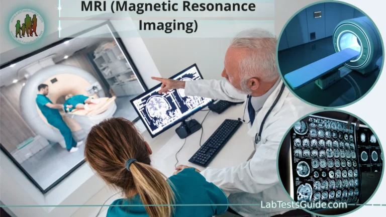
| MRI (Magnetic Resonance Imaging) | Soft tissue imaging, neurological conditions, musculoskeletal disorders, vascular imaging | No ionizing radiation, excellent soft tissue contrast, suitable for detailed brain and spine imaging. |
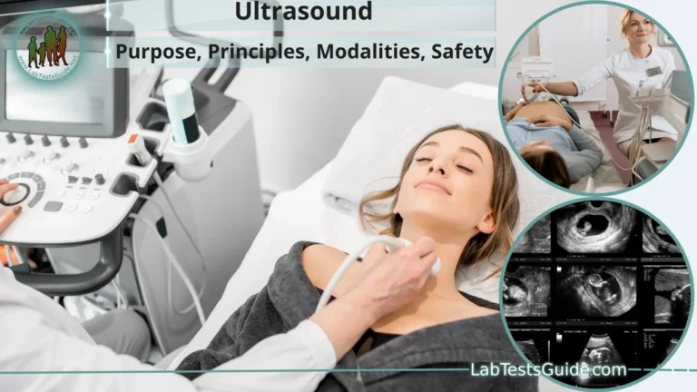
| Ultrasound (Sonography) | Obstetrics, evaluating abdominal organs, assessing blood flow, musculoskeletal injuries | No ionizing radiation, real-time imaging, safe for pregnancy, cost-effective. |
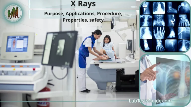
| X-ray Radiography | Bone fractures, dental imaging, chest X-rays | Quick and widely available, lower radiation dose compared to CT for certain applications, low cost. |
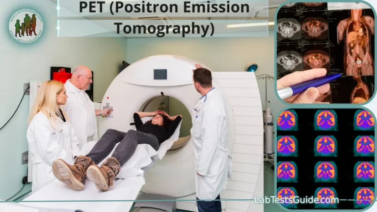
| PET (Positron Emission Tomography) Scan | Cancer staging, metabolic activity assessment, brain disorders | Reveals metabolic activity, often used in combination with CT for more comprehensive imaging. |
| Nuclear Medicine Scans (e.g., SPECT) | Assessing organ function, bone scans, cardiac perfusion imaging | Provides functional information, helps diagnose certain diseases and disorders. |
| Fluoroscopy | Real-time imaging of internal organs during procedures (e.g., barium studies, angiography) | Real-time visualization during procedures, dynamic assessment. |
| Mammography | Breast cancer screening and diagnosis | Specialized for breast imaging, high sensitivity for early breast cancer detection. |
| DEXA Scan (Dual-Energy X-ray Absorptiometry) | Bone density assessment (osteoporosis screening) | Highly accurate for measuring bone density, minimal radiation exposure. |
| Endoscopy (e.g., Colonoscopy) | Direct visualization of the gastrointestinal tract | Allows for biopsy and direct examination of tissues, essential for cancer screening and diagnosis. |
| Intravascular Ultrasound (IVUS) | Imaging blood vessels from within during cardiac procedures | Provides high-resolution images of blood vessel walls for precise diagnosis and treatment guidance. |
| Functional MRI (fMRI) | Brain mapping, assessing brain function | Maps brain activity by measuring changes in blood flow, essential for brain surgery planning. |
Future Developments in CT Imaging:
The field of CT (Computed Tomography) imaging continues to advance rapidly, driven by technological innovations and increased demands for improved diagnostic capabilities, reduced radiation exposure, and enhanced patient comfort. Here are some potential future developments in CT imaging:
- Reduced Radiation Dose: Researchers are continually working on techniques to further reduce radiation exposure in CT scans. This includes optimizing scan protocols, improving detector sensitivity, and implementing iterative reconstruction algorithms to maintain image quality while using lower radiation doses.
- Dual-Energy CT: Dual-energy CT scans use two different X-ray energy levels to provide additional information about tissue composition and material density. This technology has the potential to improve tissue characterization, differentiate between types of lesions, and enhance the detection of certain abnormalities.
- Photon-Counting Detectors: Photon-counting detectors are more sensitive and efficient than traditional detectors. They offer improved image quality, lower radiation dose requirements, and the ability to acquire high-resolution images even at low radiation levels.
- Artificial Intelligence (AI) Integration: AI and machine learning algorithms are increasingly being integrated into CT imaging to assist radiologists in image interpretation, automated lesion detection, and dose optimization. These technologies have the potential to improve accuracy and efficiency in diagnosing and reporting abnormalities.
- Functional CT Imaging: Advancements in functional CT techniques, such as perfusion imaging and CT angiography, are making it possible to assess tissue blood flow, oxygenation, and metabolic activity. This is valuable for diagnosing diseases and monitoring treatment responses.
- Cardiac CT: Cardiac CT is evolving to provide improved coronary artery visualization and assessment of cardiac function. Dual-source and high-pitch CT scanners allow for high-speed imaging, reducing the need for beta-blockers to slow heart rate.
- Cone-Beam CT: Cone-beam CT technology, commonly used in interventional radiology and radiation therapy, is becoming more widespread in diagnostic imaging. It offers 3D imaging capabilities with a lower radiation dose.
- Portable and Point-of-Care CT: The development of portable and point-of-care CT scanners is expanding access to imaging in emergency departments, intensive care units, and remote or resource-limited settings.
- Improved Image Reconstruction Algorithms: Advances in image reconstruction techniques, including model-based iterative reconstruction, promise to enhance image quality and reduce noise, especially in low-dose scans.
- Dynamic Imaging: Continuous and real-time CT scanning for dynamic applications, such as studying organ function or guiding interventions, is gaining importance in fields like interventional radiology and surgery.
- Patient-Centered Design: Future CT scanners are likely to prioritize patient comfort, with features such as wider gantries, shorter scan times, and reduced noise levels.
- Customized Scanning Protocols: AI-driven software can tailor CT scan protocols to individual patients, optimizing image quality while minimizing radiation exposure based on the specific clinical question and patient characteristics.
- Hybrid Imaging: Combining CT with other imaging modalities like PET or MRI can provide a more comprehensive assessment, particularly for cancer diagnosis, staging, and treatment planning.
FAQs:
1. What is a CT scan?
- A CT (Computed Tomography) scan is a medical imaging procedure that uses X-rays and computer technology to create detailed cross-sectional images of the body’s internal structures.
2. How does a CT scan work?
- During a CT scan, X-ray beams pass through the body from multiple angles, and detectors capture the X-rays. A computer processes this information to create detailed cross-sectional images.
3. When is a CT scan recommended?
- CT scans are recommended for diagnosing and monitoring various medical conditions, including injuries, tumors, infections, and structural abnormalities.
4. Are CT scans safe?
- While CT scans involve ionizing radiation, they are generally safe. Modern CT scanners are designed to minimize radiation exposure. The benefits of accurate diagnosis often outweigh the minimal risks.
5. How long does a CT scan take?
- The duration of a CT scan varies depending on the type of scan and the area being examined. Most scans take only a few minutes, while more complex scans may take longer.
6. Do I need to prepare for a CT scan?
- Depending on the type of CT scan, you may be asked to follow specific preparation instructions, such as fasting or discontinuing certain medications. Follow your healthcare provider’s guidance.
7. What is contrast media, and when is it used in CT scans?
- Contrast media, also known as contrast agents or contrast dyes, are substances used to enhance the visibility of certain tissues or structures in CT scans. They may be used intravenously, orally, or rectally to improve image quality.
8. Can I eat or drink before a CT scan?
- Whether you can eat or drink before a CT scan depends on the type of scan and the specific instructions provided. In some cases, fasting may be required.
9. What should I expect during a CT scan?
- During a CT scan, you will lie on a motorized table that moves through the scanner. You may be asked to hold your breath briefly during the scan. The process is painless and non-invasive.
10. Are there any side effects of a CT scan?
- Most patients experience no side effects from a CT scan. If a contrast agent is used, some may experience a warm sensation or metallic taste. Allergic reactions are rare but possible.
11. Can pregnant women have CT scans?
- Pregnant women should inform their healthcare provider and the radiology team if a CT scan is recommended. Ionizing radiation can pose risks to the developing fetus, so alternatives may be considered.
12. How are CT scan results interpreted?
- CT scan results are interpreted by radiologists, who analyze the images and provide a detailed report to the referring healthcare provider. The report guides further diagnosis and treatment.
Conclusion:
In conclusion, CT (Computed Tomography) scans are a critical tool in modern medicine, offering detailed and cross-sectional images of the body’s internal structures. They play a vital role in diagnosing and monitoring a wide range of medical conditions, from injuries to tumors and infectious diseases. While CT scans are generally safe and highly effective, they are not without risks and limitations, such as radiation exposure and the need for contrast agents in some cases.
The field of CT imaging continues to evolve, with ongoing advancements in technology, dose reduction techniques, and the integration of artificial intelligence. These developments promise to enhance the accuracy of diagnoses, improve patient safety, and expand the range of conditions that can be effectively assessed.
Patients and healthcare providers should consider the benefits and risks of CT scans for each individual case, making informed decisions about when and how to use this valuable diagnostic tool. Additionally, open communication between patients and their healthcare teams is crucial to ensuring the best possible outcomes and providing patients with the highest standard of care.
Possible References Used


