Positron Emission Tomography, commonly known as PET, is a medical imaging technique used to visualize and measure various physiological processes within the body. PET scans are particularly valuable in diagnosing and monitoring conditions such as cancer, heart disease, and neurological disorders.

- PET Definition: PET (Positron Emission Tomography) is a medical imaging technique.
- Radiotracers: It uses radioactive substances called radiotracers.
- Metabolic Imaging: PET reveals metabolic and physiological processes in the body.
- Radiotracer Injection: Radiotracers are introduced via injection or ingestion.
- Gamma Rays: PET detects gamma rays emitted during positron-electron annihilation.
- Image Creation: These rays are used to create detailed 3D images.
- Cancer Diagnosis: PET is valuable for cancer detection and staging.
- FDG-PET: Common radiotracer for measuring glucose uptake in tumors.
- Cardiac Applications: Evaluates blood flow and perfusion in heart tissue.
- Neurological Insights: Studies brain function and detects neurological disorders.
- Psychiatry: Utilized in psychiatric research to study neurotransmitter activity.
- Infection Detection: Detects increased metabolic activity in infected areas.
- Treatment Monitoring: Assesses treatment effectiveness, especially in cancer.
- Radiation Exposure: Involves ionizing radiation, so safety precautions are vital.
- Hybrid Scanners: PET-CT and PET-MRI combine PET with other imaging modalities.
- Research Tool: Crucial in drug development and preclinical research.
- Limitations: Limited spatial resolution compared to other imaging methods.
- Patient Preparation: May require fasting and avoiding strenuous exercise.
- Pregnancy and Children: Special precautions for pregnant women and children.
- Quantitative Data: Provides quantitative information about tissue activity.
- Standardized Uptake Value (SUV): Common metric for PET quantification.
- Radiotracer Half-Life: Radiotracers decay over time, limiting imaging duration.
- Image Fusion: PET images are often fused with CT or MRI for better localization.
- Clinical Guidelines: Medical societies provide guidelines for PET usage.
- Future Developments: Ongoing research aims to expand PET’s applications.
Defination of PET:
PET (Positron Emission Tomography) is a medical imaging technique that uses radioactive tracers to create detailed 3D images of metabolic and physiological processes within the body.
Purpose of PET scan:
The primary purpose of a PET (Positron Emission Tomography) scan is to visualize and assess metabolic and physiological processes within the body. PET scans are used for several purposes, including:
- Cancer Diagnosis and Staging: PET scans can detect and locate tumors in the body. They are particularly valuable in determining the stage of cancer, whether it has spread to other areas (metastasis), and in monitoring the response to cancer treatments like chemotherapy and radiation therapy.
- Cardiac Evaluation: In cardiology, PET scans can evaluate blood flow and assess the function of the heart muscle. This helps in diagnosing coronary artery disease and determining the viability of heart tissue.
- Neurological Disorders: PET is used to study brain function and to detect abnormalities in neurological disorders such as Alzheimer’s disease, Parkinson’s disease, and epilepsy.
- Psychiatry: It plays a role in psychiatric research by studying neurotransmitter activity in the brain, aiding in the understanding and diagnosis of conditions like depression, schizophrenia, and bipolar disorder.
- Infection and Inflammation: PET scans can identify areas of increased metabolic activity, making them useful in detecting infections and inflammatory conditions in the body.
- Treatment Monitoring: PET is valuable in assessing the effectiveness of treatments, especially in cancer therapy. It helps doctors evaluate whether a treatment is working and make necessary adjustments.
- Research and Drug Development: PET is a crucial tool in medical research and drug development. It allows researchers to study various physiological processes and track the effects of experimental drugs.
- Preclinical Studies: PET is used in preclinical research with animal models to investigate diseases and test potential treatments before human trials.
Historical Development of PET:
- Discovery of Positrons (1932): The PET imaging technique is based on the detection of positrons, which are the antimatter counterparts of electrons. In 1932, Carl D. Anderson discovered the positron while conducting experiments on cosmic rays.
- Theory of Annihilation (1933): The theory of positron-electron annihilation was developed, explaining how positrons interact with electrons to produce gamma rays.
- Development of Coincidence Detection (1950s): The concept of coincidence detection, essential for PET imaging, was proposed by physicists David Kuhl and Roy Edwards in the 1950s. This concept involves detecting two gamma rays produced by positron-electron annihilation simultaneously.
- First PET Scanner (1950s): Physicists Gordon Brownell and David Kuhl built the first PET scanner in the late 1950s at the University of Pennsylvania. It was a rudimentary device that could only produce two-dimensional images.
- 1970s: The 1970s saw significant developments in PET technology. Scientists like Michael Ter-Pogossian and Michael Phelps contributed to the advancement of PET scanners and radiotracers.
- Introduction of 2D PET (1970s): The introduction of 2D (two-dimensional) PET imaging allowed for better visualization of organ structures and physiological processes.
- Emergence of FDG (1970s): The radiotracer FDG (Fluorodeoxyglucose) gained prominence in the 1970s. FDG-PET became a standard in oncology for measuring glucose metabolism in tissues, aiding in cancer diagnosis and staging.
- Introduction of 3D PET (1980s): The 1980s saw the transition from 2D to 3D PET imaging, which greatly improved image quality and diagnostic accuracy.
- Hybrid Imaging (1990s): The integration of PET with other imaging modalities, such as CT (Computed Tomography) and MRI (Magnetic Resonance Imaging), led to the development of hybrid scanners like PET-CT and PET-MRI. These hybrids offered both structural and functional information in a single examination.
- Clinical Applications (1990s-Present): PET imaging found widespread clinical applications in various fields, including oncology, cardiology, neurology, and psychiatry. It became a valuable tool for diagnosing and monitoring diseases.
- Advancements in Radiotracers: Over the years, numerous radiotracers have been developed to target specific biological processes, expanding the range of PET applications.
- Research and Innovation (Present): PET technology continues to advance with ongoing research, leading to improved image quality, reduced radiation exposure, and new radiotracers for studying various physiological processes.
Principles of PET Imaging:
- Radioactive Tracers (Radiotracers): PET imaging begins with the use of radioactive substances called radiotracers. These radiotracers are typically produced by incorporating a short-lived positron-emitting radioisotope into a biologically relevant molecule. Commonly used radiotracers include FDG (Fluorodeoxyglucose), which is a radioactive form of glucose.
- Injection or Administration: Radiotracers are introduced into the patient’s body, usually through injection into a vein (intravenous injection). In some cases, they may be ingested or inhaled, depending on the specific application.
- Positron Emission: Once inside the body, the radiotracers undergo radioactive decay. During this process, the radioactive isotope emits positrons, which are positively charged subatomic particles. Positrons are the antimatter counterparts of electrons.
- Positron-Electron Annihilation: Positrons are short-lived and quickly interact with electrons within the body’s tissues. When a positron and an electron collide, they annihilate each other. This annihilation process results in the simultaneous release of two high-energy gamma-ray photons (511 keV each).
- Gamma Ray Detection: PET scanners consist of a ring of gamma-ray detectors that surround the patient. These detectors are sensitive to the gamma rays produced during positron-electron annihilation. The detectors capture the precise location and timing of each gamma ray.
- Coincidence Detection: PET scanners use a technique called “coincidence detection” to identify pairs of gamma rays that are produced nearly simultaneously. This information is crucial for creating detailed images.
- Image Reconstruction: Computer algorithms process the data from the detectors to reconstruct three-dimensional images of the distribution of radiotracers within the body. These images represent areas of high radiotracer concentration, indicating regions of increased metabolic or physiological activity.
- Quantitative Data: PET provides quantitative data, often expressed in standardized uptake values (SUVs), which measure the concentration of radiotracer in specific tissues. This quantitative information is valuable for diagnosing and monitoring diseases.
- Image Fusion: PET images are often combined or fused with images from other imaging modalities, such as CT (Computed Tomography) or MRI (Magnetic Resonance Imaging). This fusion provides both structural and functional information in a single examination, enhancing diagnostic accuracy.
Application of PET imaging:
Positron Emission Tomography (PET) imaging has a wide range of applications in medicine and medical research due to its ability to provide detailed information about metabolic and physiological processes within the body. Here are some of the key applications of PET imaging:
- Oncology (Cancer Imaging):
- PET is widely used in oncology to detect, stage, and monitor various types of cancer.
- It can identify the presence and location of tumors.
- PET is valuable in determining the extent of cancer spread (metastasis).
- It helps assess the effectiveness of cancer treatments like chemotherapy and radiation therapy.
- Cardiology (Heart Imaging):
- PET can assess blood flow to the heart muscle.
- It helps diagnose coronary artery disease (CAD) by detecting reduced perfusion.
- PET is used to evaluate the viability of heart tissue in patients with heart disease.
- Neurology (Brain Imaging):
- PET is crucial in studying brain function and neurologic disorders.
- It can detect abnormalities in conditions like Alzheimer’s disease, Parkinson’s disease, and epilepsy.
- PET helps locate regions of abnormal brain activity.
- Psychiatry (Neurotransmitter Research):
- In psychiatry, PET is used to study neurotransmitter activity in the brain.
- It aids in understanding and diagnosing conditions like depression, schizophrenia, and bipolar disorder.
- Infection and Inflammation Imaging:
- PET can identify areas of increased metabolic activity in the body.
- It helps detect infections, inflammation, and abscesses.
- Treatment Monitoring:
- PET is used to assess the response to various treatments, including cancer therapy.
- It helps clinicians determine whether a treatment is effective and make necessary adjustments.
- Research and Drug Development:
- PET plays a crucial role in medical research and drug development.
- Researchers use PET to study physiological processes, disease mechanisms, and the effects of experimental drugs.
- Preclinical Studies:
- PET is employed in preclinical research with animal models.
- It allows researchers to investigate diseases and test potential treatments before human trials.
- Neuroscience Research:
- PET is used in neuroscience to study brain function and connectivity.
- It helps researchers gain insights into cognition, behavior, and neurological disorders.
- Metabolic Studies:
- PET is used to study metabolic processes in various organs, including the liver and kidneys.
- It aids in understanding metabolic diseases and organ function.
- Pediatric Imaging:
- In pediatric medicine, PET is used to diagnose and monitor conditions like pediatric cancers, epilepsy, and developmental disorders.
- Evaluation of Cardiac Viability:
- PET helps determine whether specific regions of heart tissue are viable or scarred, guiding treatment decisions.
- Early Disease Detection:
- PET is valuable for early detection of diseases when structural changes may not yet be apparent.
- Theranostics:
- In some cases, PET is used to guide targeted therapies by identifying specific receptors or pathways that can be exploited for treatment.
- Quantitative Imaging:
- PET provides quantitative data, allowing for precise measurement of radiotracer uptake in tissues.
PET Machine Components:
- Patient Bed: This is where the patient lies during the scan. The bed can move in and out of the PET scanner to position the patient properly.
- Ring of Detectors: The core of the PET scanner consists of a ring of detectors that surrounds the patient. These detectors are sensitive to gamma rays produced during positron-electron annihilation.
- Radioisotope Delivery System: This component is responsible for delivering the radiotracer into the patient’s body. It can be a system for intravenous injection, ingestion, or inhalation, depending on the radiotracer and application.
- Collimator: Some PET scanners include a collimator, which helps control the direction of gamma rays detected. It can improve image resolution by limiting the angles at which gamma rays are detected.
- Electronic Data Acquisition System: This system collects data from the detectors. It records the precise timing and location of each gamma ray event, which is crucial for image reconstruction.
- Image Reconstruction Computer: Specialized computer algorithms process the data collected by the detectors to create three-dimensional images. These algorithms use the information about the timing and location of gamma ray events to reconstruct the radiotracer distribution within the body.
- Display and Control Console: The operator uses this console to control the PET scan, monitor the process, and review the images in real-time. It allows for adjustments and ensures the scan is proceeding as intended.
- Patient Monitoring Equipment: Vital signs, such as heart rate and oxygen saturation, are often monitored during the scan to ensure patient safety and well-being.
- Radiotracer Storage and Shielding: Radioactive radiotracers must be stored safely in lead-lined containers to prevent radiation exposure to the surroundings.
- Injection Control System: In the case of intravenous injection, an automated system can precisely control the rate and timing of the radiotracer injection.
- Motion Control System: To reduce motion artifacts in images, some PET scanners are equipped with a motion control system that compensates for patient movement during the scan.
- Gantry: The gantry is the housing that surrounds the detectors and the patient. It’s designed to shield the detectors from external sources of radiation and to maintain a stable environment for the scan.
- Table or Patient Bed Movement System: This system allows the patient bed to move in and out of the PET scanner to position the patient correctly and scan different parts of the body.
- Radiation Safety Features: PET machines are equipped with safety features to minimize radiation exposure to patients and healthcare personnel.
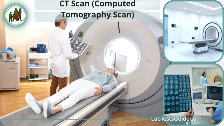
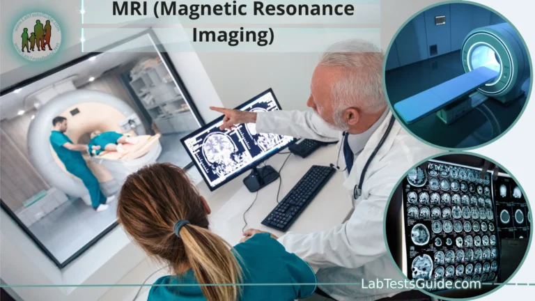
- Hybrid Imaging Capability: Some PET scanners are combined with other imaging modalities, such as CT (PET-CT) or MRI (PET-MRI), to provide both structural and functional information in a single examination.
PET Procedure and Safety:
A Positron Emission Tomography (PET) procedure is a medical imaging examination that involves the use of radioactive tracers to visualize metabolic and physiological processes within the body. Ensuring patient safety and adherence to established protocols is essential during a PET scan. Here’s an overview of the PET procedure and safety considerations:
Patient Preparation:
- Medical History and Review: Before the PET scan, the healthcare team reviews the patient’s medical history, allergies, current medications, and any existing medical conditions. It’s crucial to inform the healthcare provider about any recent surgeries or procedures.
- Fasting: Depending on the specific scan and clinical protocol, patients may be instructed to fast for a period ranging from 4 to 6 hours before the scan. Fasting helps improve the quality of images, especially in areas with high glucose metabolism, such as the abdomen.
- Medication Review: Patients should inform the healthcare team about all medications they are taking, including over-the-counter drugs and supplements. Some medications can interfere with the PET scan, and adjustments may be needed.
- Pregnancy and Breastfeeding: Female patients should inform their healthcare provider if they are pregnant or breastfeeding. In such cases, the potential risks and benefits of the PET scan will be carefully considered, and alternative imaging methods may be explored.
- Allergies: Patients should disclose any allergies, especially to contrast agents or iodine, as allergic reactions to radiotracers, though rare, can occur.
- Patient Comfort: Patients are encouraged to wear comfortable clothing and avoid clothing with metal components that may interfere with imaging. It’s important to communicate any concerns or discomfort to the healthcare team.
The PET Scan Process:
- Radiotracer Injection: The PET procedure begins with the injection of the radiotracer, which is typically administered intravenously. The radiotracer is selected based on the specific clinical purpose of the scan.
- Radiotracer Uptake Time: After the radiotracer injection, there is a period known as the “uptake time” during which the radiotracer circulates through the body and accumulates in the target tissues. This waiting period varies based on the radiotracer and clinical protocol.
- Patient Positioning: The patient lies down on the PET scanner bed, and the scan begins. Patients are instructed to remain as still as possible to ensure clear and accurate images.
Radiation Exposure and Safety:
- Radiation Exposure: PET scans involve the use of radioactive tracers, which emit positrons that interact with electrons in the body, producing gamma rays. Patients receive a dose of ionizing radiation during the scan. The radiation dose is carefully controlled and kept as low as reasonably achievable while still obtaining diagnostic-quality images.
- Radiation Monitoring: Healthcare providers and radiologic technologists are trained to minimize radiation exposure to patients and themselves. Monitoring devices are used to measure radiation levels and ensure safety.
- Pediatric Considerations: Special attention is given to pediatric patients to minimize radiation exposure. Radiotracer doses are adjusted based on the child’s size and weight.
- Radiation Safety Measures: Lead shields and collimators may be used to focus the radiation detection and minimize exposure to surrounding tissues.
- Radiation Decay: The radiotracer used in PET scans has a short half-life, which means it decays quickly and loses its radioactivity. This minimizes the duration of radiation exposure.
- Post-Procedure Hydration: Drinking plenty of fluids after the PET scan can help flush the remaining radiotracer from the body more quickly.
- Patient Monitoring: After the scan, patients are typically monitored for a brief period to ensure they are stable and not experiencing any immediate adverse reactions.
- Follow-Up: The PET images are interpreted by a radiologist or nuclear medicine physician, and the results are discussed with the patient’s healthcare provider, who will explain the findings and any recommended next steps.
Interpretation and Analysis:
1. Radiologist or Nuclear Medicine Physician Interpretation:
- The initial interpretation of PET scans is typically performed by a radiologist or a nuclear medicine physician who specializes in medical imaging.
- The interpreter reviews the PET images, paying close attention to areas of increased or decreased radiotracer uptake. These areas may indicate abnormal metabolic or physiological processes.
- They consider the patient’s medical history, clinical symptoms, and the reason for the PET scan when interpreting the images.
- The radiologist or nuclear medicine physician provides a written report summarizing their findings and impressions. This report is then shared with the referring healthcare provider.
2. Image Analysis and Quantification:
- In addition to visual interpretation, quantitative analysis may be performed to provide precise measurements of radiotracer uptake in specific regions of interest.
- Standardized uptake value (SUV) is a common metric used in PET analysis. SUV quantifies the concentration of radiotracer in a particular tissue or organ. It is calculated based on radiotracer uptake and patient body weight.
- Quantitative analysis can help in disease staging, monitoring treatment response, and assessing the extent of metabolic activity in specific areas.
3. Fusion Imaging:
- PET images are often fused with images from other modalities, such as CT (PET-CT) or MRI (PET-MRI). This fusion provides both structural and functional information in a single examination.
- The combined images help healthcare providers precisely locate areas of abnormal metabolic activity within anatomical structures.
4. Clinical Context:
- Interpretation and analysis of PET scans always take into account the clinical context. The clinical presentation of the patient, along with other diagnostic tests and medical history, is considered when arriving at a diagnosis or treatment plan.
5. Treatment Planning and Follow-Up:
- The results of PET scan interpretation play a critical role in guiding treatment decisions. For example, in oncology, PET scans help determine the stage of cancer, evaluate treatment response, and identify potential areas for biopsy or surgery.
- Follow-up PET scans may be conducted to monitor disease progression or assess the effectiveness of treatments.
6. Multidisciplinary Collaboration:
- In complex cases, a multidisciplinary team of specialists, including radiologists, nuclear medicine physicians, oncologists, surgeons, and other healthcare professionals, may collaborate to analyze PET scan results and develop a comprehensive care plan.
- This collaborative approach ensures that patients receive the most accurate diagnosis and appropriate treatment options.
Advantages and Limitations:
Positron Emission Tomography (PET) imaging is a valuable medical tool with several advantages and limitations. Here’s a breakdown of the key advantages and limitations of PET:
Advantages:
- Metabolic and Functional Imaging: PET provides information about metabolic and physiological processes within the body, offering insights into how tissues and organs are functioning. This is in contrast to anatomical imaging techniques like CT and MRI, which primarily show structures.
- Early Disease Detection: PET can detect diseases at an earlier stage than some other imaging modalities. It can identify metabolic changes before structural abnormalities become visible.
- Cancer Diagnosis and Staging: PET is highly effective in cancer diagnosis and staging. It can locate primary tumors, assess the extent of metastasis, and help guide treatment decisions.
- Treatment Monitoring: PET is valuable for monitoring the effectiveness of cancer treatments, such as chemotherapy and radiation therapy. Changes in metabolic activity can indicate treatment response or resistance.
- Neurological Disorders: PET is used to study brain function and detect abnormalities in conditions like Alzheimer’s disease, Parkinson’s disease, and epilepsy.
- Psychiatric Research: It aids in psychiatric research by studying neurotransmitter activity in the brain, contributing to the understanding and diagnosis of mental health conditions.
- Infection and Inflammation Imaging: PET can detect areas of increased metabolic activity, helping diagnose infections, inflammation, and certain immune disorders.
- Quantitative Data: PET provides quantitative data, allowing for precise measurement of radiotracer uptake in tissues. This data can be used for research and to track disease progression or response to treatment.
- Hybrid Imaging: PET can be combined with other imaging modalities like CT (PET-CT) or MRI (PET-MRI) to provide both structural and functional information in a single examination, enhancing diagnostic accuracy.
Limitations:
- Radiation Exposure: PET scans involve the use of ionizing radiation, which exposes the patient to a small amount of radiation. While the doses are generally considered safe, they should be minimized, especially in pediatric and pregnant patients.
- Limited Spatial Resolution: PET has lower spatial resolution compared to some other imaging techniques like CT and MRI. It may not provide detailed anatomical information.
- Cost: PET scans can be expensive, which can limit their availability and accessibility in some healthcare settings.
- Radiotracer Availability: Availability of specific radiotracers can be limited, and the production of some radiotracers requires specialized facilities.
- Patient Preparation: Some PET scans require fasting and specific preparations, which can be inconvenient for patients.
- False Positives and Negatives: Interpretation of PET images may be challenging, leading to false positives or negatives in some cases.
- Motion Artifacts: Patient movement during the scan can result in motion artifacts that affect image quality.
- Limited Temporal Resolution: PET may not capture rapid physiological changes due to its limited temporal resolution.
- Subject to Physiological Variability: Physiological factors such as blood flow and tissue metabolism can vary, affecting the accuracy of PET measurements.
Future Trends and Developments in PET:
The field of Positron Emission Tomography (PET) imaging continues to evolve with ongoing research and technological advancements. Several future trends and developments are expected to shape the future of PET. Here are some key areas of advancement:
- Improved Radiotracers: Researchers are working on developing new radiotracers that target specific disease processes and biological markers. This will expand the range of conditions that can be effectively studied using PET.
- Enhanced Quantitative Imaging: Advances in PET technology will lead to improved quantitative imaging capabilities, allowing for more accurate and precise measurements of radiotracer uptake. This will be particularly beneficial for research and treatment monitoring.
- Reduced Radiation Exposure: Efforts are ongoing to reduce the radiation dose associated with PET scans, making them even safer for patients, especially in pediatric and repeated scanning scenarios.
- Artificial Intelligence (AI): AI and machine learning algorithms are being developed to assist in image analysis and interpretation. These algorithms can improve the accuracy and efficiency of PET image processing and diagnosis.
- Hybrid Imaging: PET will continue to be integrated with other imaging modalities, such as CT and MRI, to provide comprehensive structural and functional information in a single examination. This hybrid imaging approach will become more routine in clinical practice.
- Personalized Medicine: PET will play a vital role in the era of personalized medicine by helping to tailor treatments to individual patients. It will assist in identifying the most effective therapies and predicting treatment responses.
- Theranostics: Theranostics combines diagnostic and therapeutic capabilities. PET will be increasingly used to guide targeted therapies by identifying specific receptors or pathways for treatment, such as in radionuclide therapy.
- Compact and Mobile PET Scanners: Advances in technology will lead to the development of smaller, more portable PET scanners. These compact systems could be used in various clinical settings, including remote and underserved areas.
- PET-MRI Integration: PET-MRI hybrid scanners will continue to improve, offering simultaneous imaging of both metabolic activity and tissue structure with high soft-tissue contrast.
- Neurological Research: PET will play a crucial role in advancing our understanding of neurological disorders, potentially leading to better diagnostics and treatments for conditions like Alzheimer’s disease and traumatic brain injuries.
- Cardiac Imaging: PET will continue to be a valuable tool in cardiology, offering insights into cardiac function, perfusion, and viability. It will aid in the assessment of heart disease and guide treatment decisions.
- Multimodal Imaging: The integration of PET with other imaging modalities, such as SPECT (Single Photon Emission Computed Tomography), will expand research and diagnostic capabilities.
- Preclinical Research: PET will remain a fundamental tool in preclinical research, enabling scientists to study diseases and test new drugs in animal models before human trials.
- Global Access: Efforts will be made to improve global access to PET technology, making it more widely available in regions where it is currently limited.
FAQs:
1. What is PET imaging?
PET (Positron Emission Tomography) is a medical imaging technique that uses radioactive tracers to create detailed 3D images of metabolic and physiological processes within the body.
2. How does PET work?
PET relies on the detection of gamma rays produced during positron-electron annihilation. Radioactive tracers emit positrons that interact with electrons in the body, resulting in gamma ray emission. PET scanners detect these gamma rays to create images.
3. What are radiotracers in PET imaging?
Radiotracers are radioactive substances used in PET imaging. They are typically combined with biologically relevant molecules and injected into the body. Radiotracers emit positrons, which are detected during the scan.
4. What is the purpose of a PET scan?
The primary purpose of a PET scan is to visualize and assess metabolic and physiological processes within the body. It is used for diagnosing, staging, and monitoring various medical conditions, including cancer, heart disease, and neurological disorders.
5. Is PET safe?
PET scans are generally considered safe. The radiation exposure is carefully controlled and kept as low as reasonably achievable. Patients should inform healthcare providers if they are pregnant or breastfeeding, as precautions may be necessary.
6. How long does a PET scan take?
The duration of a PET scan varies depending on the specific type of scan and the area being examined. It typically ranges from 20 minutes to an hour.
7. Do I need to prepare for a PET scan?
Preparation for a PET scan may include fasting for a specified period before the scan, discontinuing certain medications, and avoiding strenuous physical activity. Patients should follow the specific instructions provided by their healthcare provider.
8. What can a PET scan diagnose?
PET scans can diagnose and assess a wide range of medical conditions, including cancer, heart disease, neurological disorders, infections, and inflammatory diseases.
9. Are there any side effects of a PET scan?
PET scans are generally well-tolerated. Some patients may experience mild discomfort during radiotracer injection, and there may be rare allergic reactions. However, serious side effects are extremely rare.
10. How are PET images interpreted?
PET images are interpreted by radiologists or nuclear medicine physicians who analyze areas of increased or decreased radiotracer uptake. Quantitative measurements, such as SUV (Standardized Uptake Value), are often used for more precise analysis.
11. Is PET used in research and drug development?
Yes, PET is a valuable tool in medical research and drug development. It allows researchers to study physiological processes, disease mechanisms, and the effects of experimental drugs in preclinical and clinical studies.
12. What is the difference between PET and PET-CT or PET-MRI?
PET-CT combines PET with computed tomography (CT) to provide both metabolic and structural information. PET-MRI combines PET with magnetic resonance imaging (MRI) for enhanced soft tissue contrast and functional imaging.
Conclusion:
In conclusion, Positron Emission Tomography (PET) imaging is a powerful medical tool that allows for the visualization of metabolic and physiological processes within the human body. This non-invasive imaging technique has a wide range of applications, including the diagnosis, staging, and monitoring of various medical conditions, such as cancer, heart disease, and neurological disorders.
The principles of PET imaging, which involve the detection of gamma rays produced during positron-electron annihilation, enable the creation of detailed 3D images. These images provide valuable insights into the function and activity of tissues and organs, complementing other imaging modalities like CT and MRI.
Possible References Used


