Blood agar is a type of solid growth medium used in microbiology laboratories to culture and identify bacteria. It is composed of a nutrient-rich agar base supplemented with sterile blood, typically sheep or horse blood. The blood serves as an enriching agent and provides essential nutrients for bacterial growth.
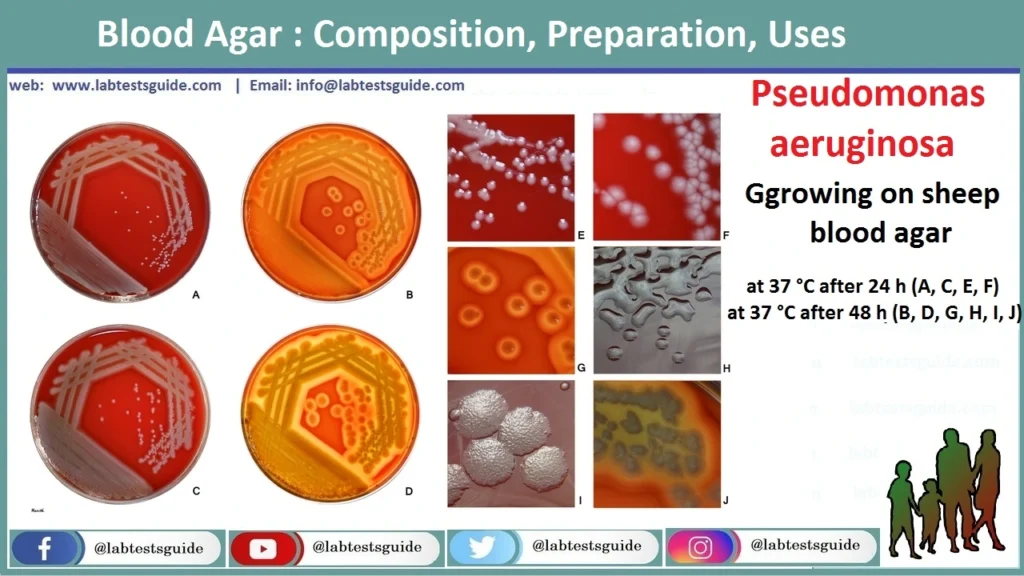
Examples of bacteria that grow in blood agar medium.
- Haemophilus influenza
- Neisseria species
- Strep Pneumonia
Agar blood as a differential medium.
Blood agar is a differential medium because it helps detect and differentiate hemolytic bacteria such as Streptococcus species. Detects hemolysis by cytolytic toxins that are secreted by a strain of bacteria.
Agar blood as a selective medium.
Blood agar can be used as a selective medium for some types of pathogens. To be selective, you must add a chemical, a dye or an antibiotic.
Blood agar Components
- Agar
- Pancreatic digest of casein
- Sodium chloride
- Papaic digest of soy meal
- Beef extract
- Peptone
- Sheep blood, defibrinated
- Distilled water
- pH 7.3 ± 0.2 at 25°C
For 1 Liter Preparation
| Items | Volume |
| Agar | 15.0 g |
| Beef extract | 10.0 g |
| Peptone | 10.0 g |
| Sodium Chloride (NaCl) | 5,0 g |
| Sheep blood, defibrinated | 50 ml |
| pH | 7.3 ± 0.2 at 25°C |
| Distilled/Deionized | 950 ml |
Blood agar Preparation
- Add above components (40 gm), except sheep blood, to distilled/deionized water and bring volume to 950.0 mL.
- Mix thoroughly.
- Heat with frequent agitation and boil for 1 min to completely dissolve.
- Autoclave for 15 min at 15 psi pressure at 121°C.
- Cool to 45°- 50°C.
- Aseptically add 50.0 mL of sterile, defibrinated sheep blood.
- Mix thoroughly and pour into sterile Petri dishes.
What is blood agar used for?
- It is used to isolate and identify antimicrobial susceptibility of Streptococci.
- It is used to isolate and cultivate Streptococci, Neisseria, and fastidious microorganisms.
- Blood agar helps differentiate bacteria based on their haemolytic properties.
- It is used to prepare Salmonella typhi antigens.
- It determines the salinity range of marine Flavobacteria.
- Blood agar base can be used for testing food samples as per the recommendation of APHA.
- It helps determine the type of hemolysis.
What Bacteria grow on blood agar?
- Neisseria
- Haemophilus genera
- Streptococcus pyogenes
- S. pneumonia
- Satellitism of H. influenza
- Streptococcus pneumonia
- Streptococci-Streptococcus agalactiace
- Clostridium perfringens
Result Interpretation on Blood Agar
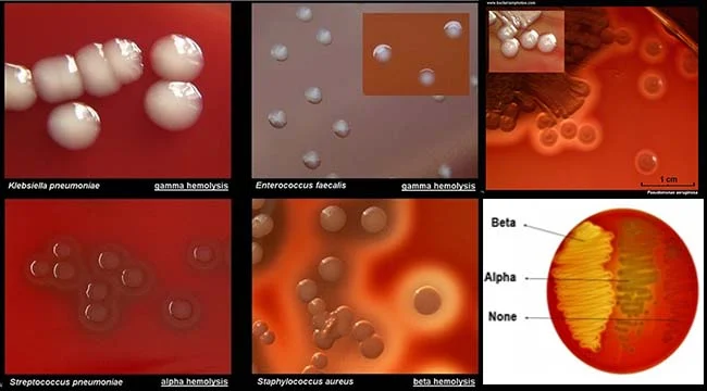
The basal medium appears light amber colored which might look clear to slightly opalescent gel. After the addition of 5%, v/v sterile defibrinated blood; however, the cherry red-colored opaque gel is formed on the Petri plates. The following table demonstrates the growth of important medical bacteria with their colony morphologies on Blood Agar Medium:
| Sr # | Organism | Growth | Colony Morphology | Hemolysis |
|---|---|---|---|---|
| 1 | Neisseria meningiditis | Good-luxuriant | Grey and unpigmented colonies that appear round, smooth, moist, glistening, and convex, with a clearly defined edge. | Non-hemolytic or γ-hemolytic. |
| 2. | Salmonella Typhi | Good-luxuriant | Smooth colorless colonies that are smooth, moist, and flat with the diameter range of 2-4 mm. | Non-hemolytic or γ-hemolytic. |
| 3. | Staphylococcus aureus | Luxuriant | Golden yellow colored circular, convex and smooth colonies of the diameter range of 2-4 mm; opaque colonies with a zone of hemolysis. | β-hemolytic. |
| 4. | Staphylococcus epidermidis | Luxuriant | Circular, colonies of the size 1-4 mm in diameter; grey to white-colored with low convex elevation; moist, glistening colonies. | Non-hemolytic or γ-hemolytic. |
| 5. | Streptococcus pyogenes | Luxuriant | White-greyish-colored colonies with a diameter of > 0.5 mm; the colonies are surrounded by a zone of β-hemolysis that is often two to four times as large as the colony diameter. | β-hemolytic. |
| 6. | Streptococcus pneumonia | Luxuriant | small, grey, moist (sometimes mucoidal in encapsulated virulent strains), colonies with the characteristic zone of alpha-hemolysis (green); due to autolysis, often produces a dimple-like zone of hemolysis than the typical crater-like appearance. | α-hemolytic. |
| 7. | Pseudomonas aeruginosa | Good-luxuriant | Large colonies of the size 2-5mm in diameter; flat colonies that are grey to white-colored with an undulate margin with a zone of β-hemolysis. | β-hemolytic. |
What is the difference between blood agar and chocolate agar?
Blood agar and chocolate agar are quite similar but they differ in preparation. Blood agar consists of many ingredients but the primary ingredient is the blood, which came from a rabbit or a sheep.
It is treated and water and a number of other ingredients are added to sterilize the solution. The blood is treated to remove fibrin, the clotting factor of the blood. On the other hand, chocolate agar contains lysed red blood cells, which turns brown giving the medium its chocolate color.
Possible References Used


One Comment