Microscope Structure, Types, Functions, Parts, Diagrams and more detail
Explore the world of microscopy with information on microscope types, labeled diagrams, and detailed insights into the parts of a microscope. From the basics to advanced techniques like scanning probe microscopy, discover the diverse applications of microscopes in scientific research.
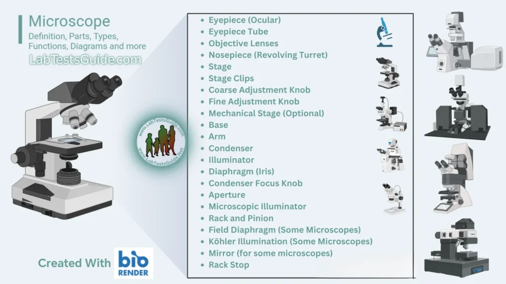
Microscopes are essential scientific instruments that magnify and illuminate tiny objects, enabling detailed observation of microscopic structures. They come in various types, each serving specific purposes, from basic optical microscopes to advanced electron and scanning probe microscopes. Dive into the microscopic world, exploring labeled diagrams and understanding the diverse components that make up these powerful tools.
Definition of Microscope:
A microscope is a scientific instrument that magnifies and allows detailed observation of small objects or structures that are not visible to the naked eye.

Microscopy in laboratories
- Malaria
- Trypanosomiasis
- Leishmaniasis
- Onchocerciasis
- Lymphatic filariasis
- Loiasis
- Schistosomiasis
- Amoebiasis
- Giardiasis
- Cryptosporidiosis
- Intestinal Worm Infections
- Paragonimiasis
- Tuberculosis
- Leprosy
- Meningitis
- Gonorrhoea (Male)
- Cholera
- Urinary Tract Infections
- Trichomoniasis
- Vaginal candidiasis
- Red cells in anaemia
- White cell changes
- Skin mycoses
History of Microscope:
- Early Optical Devices:
- The history of microscopes dates back to the late 16th century when eyeglass makers created the first crude optical devices that could magnify small objects.
- Hans Lippershey’s Contribution (1590s):
- Dutch spectacle maker Hans Lippershey is often credited with inventing the first compound microscope around 1590, using a convex and a concave lens.
- Zacharias Janssen’s Innovation (1595):
- Zacharias Janssen, another Dutchman, is attributed with the invention of the first compound microscope in 1595, using a tube with lenses at both ends.
- Antonie van Leeuwenhoek’s Single-Lens Microscopes (17th Century):
- In the mid-17th century, Antonie van Leeuwenhoek, a Dutch scientist, advanced microscopy by developing single-lens microscopes, achieving remarkable magnification and making significant biological discoveries.
- Robert Hooke’s Micrographia (1665):
- Robert Hooke’s “Micrographia” in 1665 popularized microscopy with detailed illustrations of biological specimens, including the first use of the term “cell.”
- Improvements in Optics (18th Century):
- The 18th century saw advancements in optics, leading to improved microscope lenses and enhanced magnification.
- Compound Microscope Refinement (19th Century):
- Throughout the 19th century, the compound microscope underwent refinements in design and optics, making it a standard tool in scientific research.
- Electron Microscope Development (20th Century):
- The 20th century witnessed the development of electron microscopes, starting with the transmission electron microscope (TEM) in the 1930s, followed by the scanning electron microscope (SEM) later on.
- Scanning Probe Microscopes (1980s):
- In the 1980s, the invention of scanning probe microscopes, such as the Atomic Force Microscope (AFM), introduced new techniques for imaging surfaces at the atomic level.
- Modern Advancements (21st Century):
- The 21st century continues to see advancements in microscopy, with the integration of digital technology, fluorescence microscopy techniques, and ongoing innovations in microscopy applications across various scientific disciplines.
Types of Microscope:
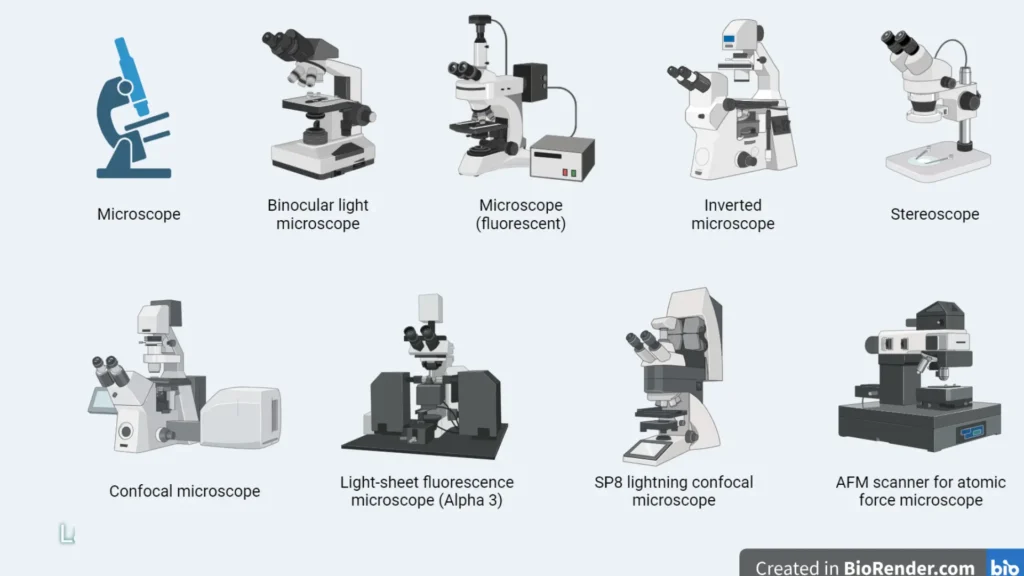
- Optical Microscopes:
- Compound Microscope: Uses multiple lenses for high magnification.
- Stereo Microscope (Dissecting Microscope): Provides a three-dimensional view for larger specimens.
- Electron Microscopes:
- Transmission Electron Microscope (TEM): Passes electrons through a specimen for high-resolution internal imaging.
- Scanning Electron Microscope (SEM): Scans a focused electron beam over a specimen for detailed surface imaging.
- Scanning Probe Microscopes:
- Atomic Force Microscope (AFM): Uses a sharp tip to scan surfaces at the atomic level.
- Scanning Tunneling Microscope (STM): Measures electron tunneling for atomic-scale imaging.
- Fluorescence Microscopes:
- Widefield Fluorescence Microscope: Illuminates and detects fluorescent signals.
- Confocal Laser Scanning Microscope (CLSM): Uses lasers to create high-contrast images.
- Digital Microscopes:
- Digital Light Microscope: Incorporates digital cameras for image capture.
- Digital Electron Microscope: Utilizes digital technology for electron microscopy.
- Contrast-Enhancing Microscopes:
- Phase-Contrast Microscope: Enhances contrast in transparent specimens.
- Differential Interference Contrast Microscope (DIC): Emphasizes variations in specimen thickness.
- Darkfield Microscope: Illuminates specimens against a dark background for enhanced contrast.
- Polarizing Microscope: Analyzes materials’ optical properties by polarized light.
- Inverted Microscope: Positions the objective lens below the specimen, suitable for live cell cultures.
- Acoustic Microscope: Uses sound waves for imaging, useful in materials science.
- Virtual Microscope: Utilizes digital technology for virtual specimen exploration.
- Near-Field Scanning Optical Microscope (NSOM or SNOM): Provides nanoscale optical imaging.
- Raman Microscope: Combines microscopy with Raman spectroscopy for molecular analysis.
- Super-Resolution Microscope (STED, SIM, PALM, STORM): Surpasses the diffraction limit for increased resolution.
- Ultraviolet Microscope: Uses ultraviolet light for imaging, valuable in fluorescence studies.
- X-ray Microscope: Utilizes X-rays for imaging dense materials.
- Cryo-Electron Microscope: Images specimens at cryogenic temperatures, preserving biological structures.
- Environmental Microscope (ESEM): Allows imaging in a controlled environmental chamber, suitable for wet or unprocessed samples.
Each type of microscope serves specific purposes, enabling diverse applications in scientific research and exploration.
State Functions of a Microscope:
The functions of a microscope are geared toward magnifying and illuminating tiny objects, enabling detailed observation and analysis. The specific functions may vary depending on the type of microscope, but here are general functions applicable to many types:
- Magnification:
- Enlarges the image of the specimen, allowing for a detailed examination.
- Resolution:
- Defines the clarity and detail of the image, providing the ability to distinguish between closely spaced objects.
- Illumination:
- Provides a light source to illuminate the specimen, enhancing visibility.
- Focusing:
- Allows adjustment of the focal plane to achieve a sharp and clear image.
- Stage Control:
- Enables movement of the specimen stage for positioning and scanning.
- Condenser Adjustment:
- Adjusts the condenser to control the intensity and direction of the light on the specimen.
- Objective Lenses:
- Houses different lenses with varying magnifications, providing flexibility in observation.
- Eyepiece (Ocular Lens):
- Contains lenses for further magnification and observation by the viewer.
- Coarse and Fine Adjustment:
- Coarse adjustment rapidly moves the stage for initial focusing, while fine adjustment allows precise focusing for detailed examination.
- Illumination Control:
- Adjusts the brightness or intensity of the light source for optimal specimen illumination.
- Polarizer and Analyzer (Polarizing Microscopes):
- Facilitates the analysis of materials with polarized light, revealing specific optical properties.
- Objective Turret (Nosepiece):
- Holds multiple objective lenses, enabling quick and easy switching between magnifications.
- Filter Slots (Fluorescence Microscopes):
- Accommodates filters for selecting specific wavelengths of light, crucial for fluorescence studies.
- Aperture Diaphragm:
- Controls the diameter of the light beam, influencing resolution and contrast.
- Köhler Illumination (for some microscopes):
- A method of specimen illumination that provides even and adjustable light distribution.
- Camera Port (for Digital Microscopes):
- Allows the attachment of cameras for image and video capture.
- Stage Clips or Mechanical Stage:
- Secures the specimen in place and facilitates precise movement for scanning.
- Specialized Features (depending on microscope type):
- Examples include phase-contrast optics, darkfield condensers, environmental chambers, and more, catering to specific observation needs.
The combination of these functions allows researchers, scientists, and students to explore the microscopic world and conduct detailed analyses in various scientific disciplines.
Structural Parts of Microscope:
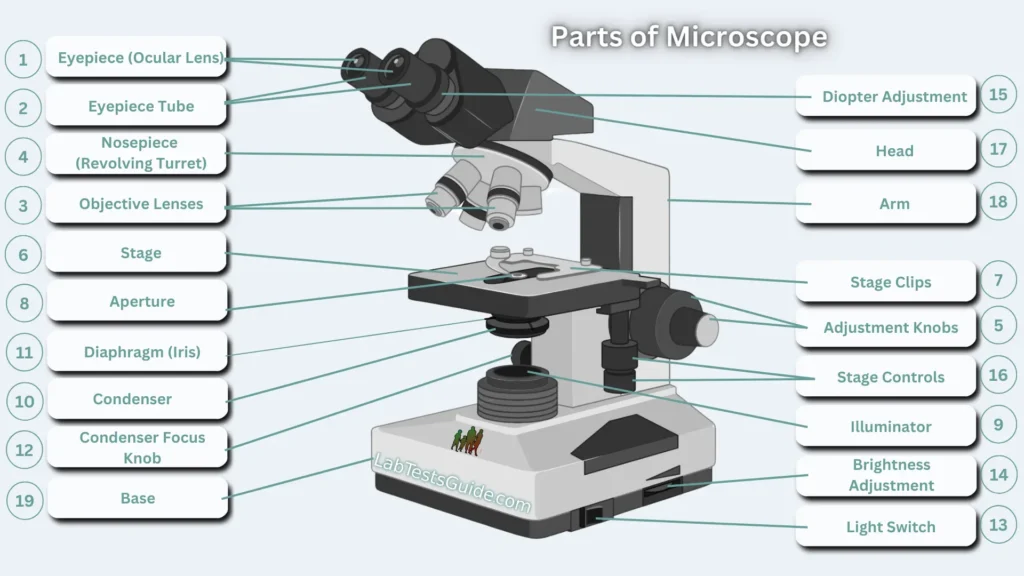
Microscopes are indispensable tools in scientific exploration, allowing us to delve into the intricate world of the unseen. To comprehend the functionality of these powerful instruments, it’s crucial to familiarize ourselves with their three main structural parts: the head, base, and arm.
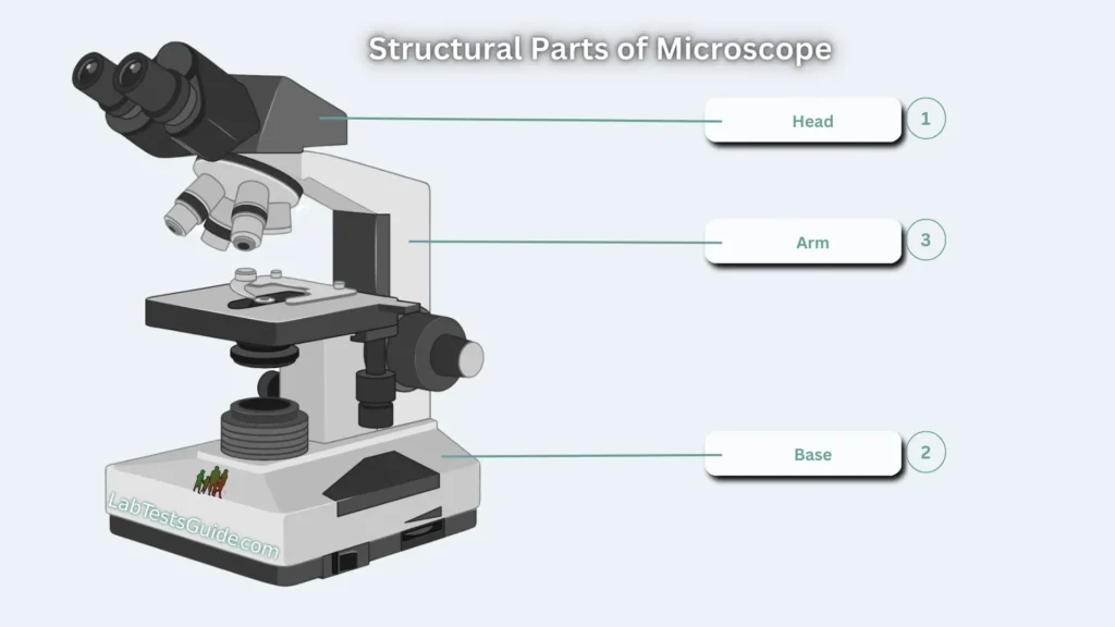
1. Head : The Command Center of Observation
The head, also known as the body, serves as the command center of the microscope. Positioned in the upper part, it houses the optical components that are vital for magnification and observation. Within the head lie the eyepiece and objective lenses, which play a crucial role in capturing and magnifying the microscopic details of specimens.
2. Base: The Pillar of Support and Illumination
Functioning as the foundational support, the base is the bottom part of the microscope. Beyond providing stability, the base often carries microscopic illuminators, ensuring proper lighting for the specimen under observation. This illuminating mechanism is essential for creating optimal conditions for detailed examination.
3. Arm: Bridging Strength and Flexibility
The arm acts as the bridge connecting the base to the head and facilitates the movement of the microscope. It provides essential support to the head, ensuring stability during observations. Notably, the arm is a crucial element when transporting the microscope. In some high-quality models, an articulated arm with multiple joints enhances the range of movement for the microscopic head, facilitating better viewing angles and flexibility during research.
Optical Parts of Microscope:
Microscopes, the gateways to the microscopic world, rely on a set of intricate optical parts to illuminate, magnify, and produce detailed images from specimens. Let’s delve into these optical components that transform the unseen into the visible:
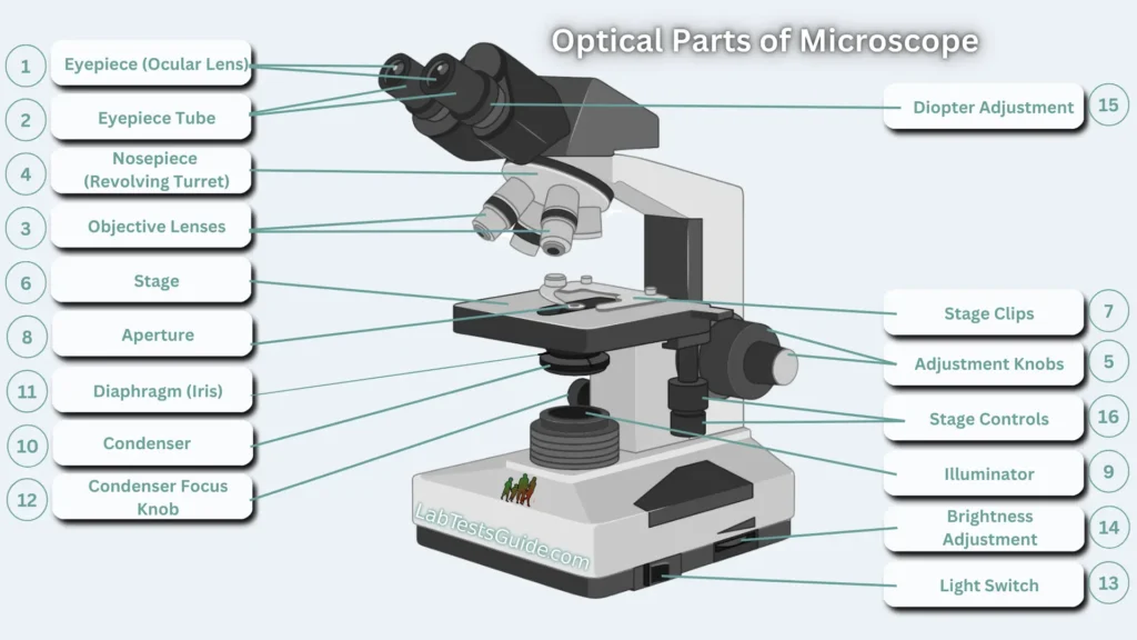
1. Eyepiece (Ocular): The Observer’s Window
- The eyepiece, or ocular, is the lens through which the viewer peers to observe the specimen. Positioned at the top of the microscope, it standardly offers 10x magnification, with optional eyepieces ranging from 5x to 30x.
2. Eyepiece Tube: Supporting Visual Insight
- The eyepiece tube holds the eyepiece in place, providing structural support for visual observation. In some microscopes, such as binocular models, the tube is flexible and rotatable for optimal visualization and adjustable distance. For monocular microscopes, the tube is typically fixed.
3. Objective Lenses: Magnification Masters
- These are the primary lenses responsible for visualizing the specimen. Ranging in magnification from 40x to 100x, microscopes typically feature 1 to 4 objective lenses. Some face forward, while others face backward, and each lens has its unique magnification power.
- Scanning Objective Lens (4x)
- Low Power Objective (10x)
- High Power Objective Lens (40x)
- Oil Immersion Objective Lens (100x)
- Specialty Objective Lenses (2x, 50x Oil, 60x and 100x Dry)
4. Nosepiece (Revolving Turret): Lenses in Rotation
- The nosepiece, also known as the revolving turret, holds multiple objective lenses. Its movable design allows for the rotation of objective lenses, enabling users to select different magnifications for observation.
5. Adjustment Knobs: Precision Focusing
- Adjustment knobs, both fine and coarse, are crucial for focusing the microscope. The coarse adjustment knob facilitates rapid movement, while the fine adjustment knob provides meticulous control, ensuring a clear and sharp image.
6. Stage: Platform for Observation
- The stage is a flat platform where the specimen is placed for viewing. It often includes stage clips to secure specimen slides. Mechanical stages, common in many microscopes, allow controlled movement using mechanical knobs.
7. Stage Clips:
- Metal clips securing the specimen slide in place on the stage.
8. Aperture: Gateway for Illumination
- The aperture is a hole on the microscope stage, serving as a pathway for transmitted light from the source to reach the specimen.
9. Microscopic Illuminator: Shedding Light on the Unseen
- Located at the base, the microscopic illuminator is the light source for the microscope. It replaces the traditional mirror and captures light from an external low-voltage source, typically around 100V.
10. Condenser: Focusing Light for Clarity
- Found under the stage, the condenser collects and focuses light from the illuminator onto the specimen. It plays a crucial role in producing clear and sharp images, especially at high magnifications of 400x and above.
11. Diaphragm (Iris): Controlling Light Intensity
- The diaphragm, also known as the iris, is located under the stage. It controls the amount of light reaching the specimen, allowing for adjustable light intensity and beam size.
12. Condenser Focus Knob: Controlling Light Focus
- This knob adjusts the position of the condenser, controlling the focus of light on the specimen.
13. Light Switch
- The light switch on a microscope controls the illumination source, allowing users to turn the light on or off as needed during observation.
14. Brightness Adjustment
- The brightness adjustment feature on a microscope regulates the intensity of the light source, providing control over the illumination level for optimal specimen visibility.
15. Diopter Adjustment:
- The diopter adjustment is a setting on the microscope eyepiece that allows users to customize the focus of the eyepiece to match their individual vision, enhancing viewing comfort and clarity.
16. Stage Controls:
- Stage controls on a microscope refer to mechanisms that allow precise manipulation and positioning of the specimen on the stage. These controls include knobs or buttons for horizontal (X-axis) and vertical (Y-axis) movement, enabling users to scan and focus on different areas of the specimen with accuracy and ease.
Microscopy Terms :
Microscopy involves a specific set of terms that describe various aspects of the tools, techniques, and observations in this field. Here are some commonly used microscopy terms:
- Magnification:
- The degree to which an object is enlarged under the microscope compared to its actual size.
- Resolution:
- The ability of a microscope to distinguish between two points that are close together. Higher resolution provides clearer and more detailed images.
- Objective Lens:
- The primary lens or set of lenses closest to the specimen, responsible for magnification.
- Eyepiece (Ocular):
- The lens or set of lenses through which the observer looks, further magnifying the image produced by the objective lens.
- Field of View:
- The area visible through the microscope when looking into the eyepiece.
- Condenser:
- A lens system that focuses light onto the specimen, improving image contrast and brightness.
- Illumination:
- The source of light used to illuminate the specimen for observation.
- Contrast:
- The difference in intensity or color between an object and its background, influencing visibility.
- Staining:
- The process of adding colored dyes to specimens to enhance contrast and highlight specific structures.
- Darkfield Microscopy:
- A technique where light is directed at an angle to the specimen, creating a dark background and emphasizing light-scattering structures.
- Fluorescence Microscopy:
- Using fluorescence to visualize specimens by exposing them to light, causing them to emit colored light.
- Phase-Contrast Microscopy:
- A technique that enhances the contrast of transparent and colorless specimens.
- Differential Interference Contrast (DIC) Microscopy:
- A method of microscopy that improves contrast by detecting variations in refractive index.
- Transmission Electron Microscopy (TEM):
- A technique that uses electrons to image the internal structures of thin specimens.
- Scanning Electron Microscopy (SEM):
- A technique that uses electrons to create detailed three-dimensional surface images of specimens.
- Atomic Force Microscopy (AFM):
- A type of scanning probe microscopy that uses a sharp tip to scan the surface of a specimen at the atomic level.
- Confocal Microscopy:
- A technique that uses lasers to create sharp, high-contrast images, often used in biological and materials research.
- Resolution Limit:
- The smallest distance between two points that can be distinguished as separate entities under a microscope.
- Numerical Aperture:
- A measure of the ability of a lens to gather light and resolve fine details, influencing image quality.
- Z-stack:
- A series of images taken at different focal planes, used to create a three-dimensional representation of a specimen.
These terms provide a foundational understanding of microscopy and are essential for researchers, scientists, and students working in the field.
Home | Blog | About Us | Contact Us | Disclaimer
Possible References Used



9 Comments