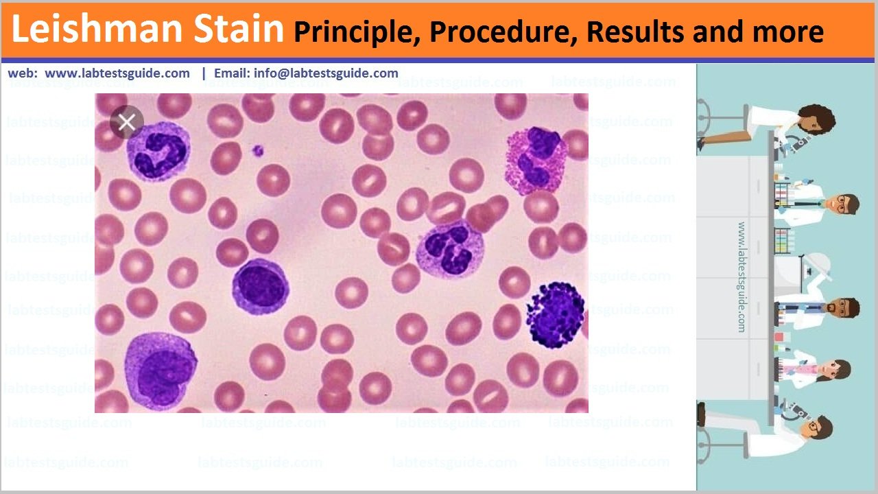
Leishman stain, also known as Leishman’s stain, is used in microscopy for staining blood smears. It is generally used to differentiate between and identify white blood cells, malaria parasites, and trypanosomas.

Requirements:
- Leishman Stain (Stock Solution)
- Microscopic Glass Slide
- Phosphate buffer (pH 6.8)
- Graduated pipettes
- Measuring cylinder
- Distilled Water
- Pasteur pipette
- Coplin Jar
- Blood Specimen
Principle:
Leishman stain, is used in microscopy for staining blood smears. It provides excellent stain quality. It is generally used to differentiate and identify leucocytes, malaria parasites, and trypanosomas. It is based on a methanolic mixture of “polychromed” methylene blue. (i.e. demethylated into various azures) and eosin.The methanolic stock solution is stable and also serves the purpose of directly fixing the smear eliminating a prefixing step.
Ingredients:
- Leishman stain – 0.150 gm
- Methanol, absolute – 100.000 ml
Procedure:
- Use smears that are as thin as possible and air-dried.
- Fully cover the smears with Leishman’s Stain solution. Stain for 2 minuted.
- Add twice the amount of distilled water and mix by swirling. Incubate for at least 10 min.
- Rinse thoroughly with distilled water.
- Dry the slides using blotting paper and air-dry
The color of Nuclei by Leishman Stain
| Chromatin | Purple |
| Nucleoli | Light blue |
The color of Cytoplasm by Leishman Stain
| Erythrocytes | Pink |
| Reticulocytes | Dark blue |
| Lymphocytes | Blue |
| Monocytes | Grey blue |
| Neutrophils | Bluish Pink |
| Basophils | Blue |
The color of Granules by Leishman Stain
| Basophil | Purple black |
| Eosinophil | Red orange |
| Neutrophil | Purple |
| Platelet | Purple |
Related Articles:
Possible References Used


